Novus Biologicals products are now on bio-techne.com

LYVE-1 Antibody - BSA Free

Images

 
Western Blot: LYVE-1 Antibody [NB100-725] - Western Blot: [NB100-725] - Total protein from human stomach, lymph node and placenta was separated on a 7.5% gel by SDS-PAGE, transferred to PVDF membrane and blocked in 5% ...read more
Immunohistochemistry-Paraffin: LYVE-1 Antibody [NB100-725] - Analysis using the DyLight 488 conjugate of NB100-725. Staining of LYVE1 in paraffin embedded MDA-MB-231 breast cancer orthotopic transplantation tissue. ...read more
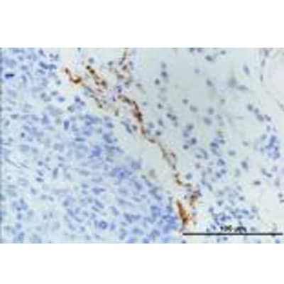
Immunohistochemistry: LYVE-1 Antibody [NB100-725] - Detection of LYVE1 in endothelial cells of human lung blood vessels using NB100-725. Note presence of RBCs within vessel lumen.
Flow Cytometry: LYVE-1 Antibody [NB100-725] - Analysis using the DyLight 488 conjugate of NB100-725. Unlabeled control with labeled samples of cells dissociated mechanically and enzymatically from human skin. Image ...read more
Western Blot: LYVE-1 Antibody [NB100-725] - Analysis of extracts from A549 cells using LYVE1 antibody (NB100-725, 1:100). Image from verified customer review.
Flow Cytometry: LYVE-1 Antibody [NB100-725] - LYVE1 antibody was tested at 1:400 in A549 cells using an Alexa Fluor 488 secondary (shown in green) alongside unstained cells (shown in red).
Flow Cytometry: LYVE-1 Antibody [NB100-725] - An intracellular stain was performed on A549 cells with LYVE-1 Antibody NB100-725 (blue) and a matched isotype control (orange). Cells were fixed with 4% PFA and then ...read more
Immunohistochemistry: LYVE-1 Antibody [NB100-725] - Detection of LYVE1 in endothelial cells of human bladder vasculature using NB100-725.
Immunohistochemistry: LYVE-1 Antibody [NB100-725] - Detection of LYVE1 in endothelial cells of human lung blood vessels using NB100-725.
Flow Cytometry: LYVE-1 Antibody [NB100-725] - Analysis using the Alexa Fluor (R) 488 conjugate of NB100-725. Staining of LYVE1 in human dissociated skin lyve1+ using AF488 conjugated anti-LYVE1 antibody. Image from ...read more
Flow Cytometry: LYVE-1 Antibody [NB100-725] - An intracellular stain was performed on A549 cells with LYVE-1 Antibody NB100-725G (blue) and a matched isotype control (orange). Cells were fixed with 4% PFA and then ...read more
Flow Cytometry: LYVE-1 Antibody [NB100-725] - An intracellular stain was performed on K562 cells with LYVE-1 Antibody NB100-725C (blue) and a matched isotype control (orange). Cells were fixed with 4% PFA and then ...read more

Product Details

Summary
Reactivity Hu, Mu, RtSpecies Glossary
Applications WB, Flow, IHC
Clonality
Polyclonal
Host
Rabbit
Conjugate
Unconjugated
Format
BSA Free
Concentration
1.0 mg/ml

Order Details

Novus Biologicals is part of Bio-Techne

Shop this product on bio-techne.com

LYVE-1 Antibody - BSA Free Summary

Immunogen
This LYVE-1 Antibody was developed against a synthetic peptide made to a C-terminal portion of the mouse LYVE1 protein sequence (between residues 250-318). [UniProt# Q8BHC0]
Localization
Plasma membrane
Marker
Lymphatic Vessel Marker
Isotype
IgG
Clonality
Polyclonal
Host
Rabbit
Gene
LYVE1
Purity
Immunogen affinity purified
Innovator's Reward
Test in a species/application not listed above to receive a full credit towards a future purchase.

Applications/Dilutions

Dilutions
  • Flow (Intracellular)
  • Flow Cytometry 1:50-1:500
  • Immunohistochemistry 1:100-1:200
  • Immunohistochemistry-Frozen 1:100-1:200
  • Immunohistochemistry-Paraffin 1:100-1:200
  • Western Blot 1:500-1:2000
Application Notes
In Western Blot, a band at ~52 kDa is seen. For Immunohistochemistry citrate buffer antigen retrieval is recommended.
Theoretical MW
35 kDa.
Disclaimer note: The observed molecular weight of the protein may vary from the listed predicted molecular weight due to post translational modifications, post translation cleavages, relative charges, and other experimental factors.
Control
Mouse Spleen Whole Tissue Lysate (Adult Whole Normal)
Control Peptide
LYVE-1 Protein (NB100-725PEP)
Reviewed Applications
Read 2 Reviews rated 5
using
NB100-725 in the following applications:

Publications
Read Publications using
NB100-725 in the following applications:

Packaging, Storage & Formulations

Storage
Store at 4C short term. Aliquot and store at -20C long term. Avoid freeze-thaw cycles.
Buffer
PBS
Preservative
0.02% Sodium Azide
Concentration
1.0 mg/ml
Purity
Immunogen affinity purified

Alternate Names for LYVE-1 Antibody - BSA Free

  • cell surface retention sequence binding protein-1
  • Cell surface retention sequence-binding protein 1
  • CRSBP1
  • CRSBP-1
  • extracellular link domain containing 1
  • extracellular link domain-containing 1
  • Extracellular link domain-containing protein 1
  • HAR
  • Hyaluronic acid receptor
  • lymphatic vessel endothelial hyaluronan receptor 1
  • lymphatic vessel endothelial hyaluronic acid receptor 1
  • LYVE1
  • LYVE-1
  • UNQ230/PRO263
  • XLKD1

Background

LYVE-1 (Lymphatic Vessel Endothelial Receptor 1) is a receptor for the extracellular matrix mucopolysaccharide hyaluronan (HA) and is primarily expressed by lymphatic endothelial cells in addition to the sinusoidal endothelium of the liver and spleen (1-3). HA, also called hyaluronic acid, is a main component of the extracellular matrix and functions largely in cell adhesion, migration, and tissue remodeling (2). HA undergoes a turnover process that involves release from the tissues to the afferent lymph, degradation within the lymph nodes, and removal of fragments by the liver (2,3). LYVE-1, in addition to other lymphatic proteins including VEGFR3, Prox1, and podoplanin, is a common marker for differentiating between the blood and lymphatic systems (2,3). Furthermore, LYVE-1 is closely related to the leukocyte receptor CD44, having ~44% sequence similarity (1-3). Like CD44, the LYVE-1 protein contains an extracellular HA-binding link domain, with N- and C-terminal extensions, located at the end of a glycosylated juxtamembrane domain stalk region, followed by a transmembrane region, and a cytoplasmic tail (1-3). The key features of the HA-binding like molecule are the three disulfide bridges formed by six cysteine residues (1-3). LYVE-1 is synthesized as a protein of 322 amino acids in length with a theoretical molecular weight of 35 kDa, although due to O-glycosylation often appears in SDS-PAGE at a molecular weight ranging from ~60-70 kDa (2,4). The role of HA-binding is further elucidated by the identification of LYVE-1 as a docking receptor for dendritic cells and macrophages, binding their surface HA to control the entry and migration into lymph vessels (1).

LYVE-1 has been an important marker in studies of embryonic and tumor lymphangiogenesis, as many cancers are characterized by early metastasis to the lymph nodes (1-3, 5). One study of five different vascular tumors in infants used immunohistochemical analysis and found positive LYVE-1 expression in infantile hemangioma, tufted angioma, and kaposiform hemangioendothelioma (5). LYVE-1 along with other markers such as GLUT-1, CD31, CD34, Prox-1, and WT-1 can be used to help provide immunohistologic profiles of various tumors and, when used in conjunction with clinical and histopathologic approaches, may offer better overall diagnosis and disease treatment (5).

References

1. Jackson D. G. (2019). Hyaluronan in the lymphatics: The key role of the hyaluronan receptor LYVE-1 in leucocyte trafficking. Matrix Biology : Journal of the International Society for Matrix Biology. https://doi.org/10.1016/j.matbio.2018.02.001

2. Jackson D. G. (2004). Biology of the lymphatic marker LYVE-1 and applications in research into lymphatic trafficking and lymphangiogenesis. APMIS : acta pathologica, microbiologica, et immunologica Scandinavica. https://doi.org/10.1111/j.1600-0463.2004.apm11207-0811.x

3. Jackson D. G. (2003). The lymphatics revisited: new perspectives from the hyaluronan receptor LYVE-1. Trends in Cardiovascular Medicine. https://doi.org/10.1016/s1050-1738(02)00189-5

4. Unitprot (Q9Y5Y7)

5. Johnson, E. F., Davis, D. M., Tollefson, M. M., Fritchie, K., & Gibson, L. E. (2018). Vascular Tumors in Infants: Case Report and Review of Clinical, Histopathologic, and Immunohistochemical Characteristics of Infantile Hemangioma, Pyogenic Granuloma, Noninvoluting Congenital Hemangioma, Tufted Angioma, and Kaposiform Hemangioendothelioma. The American Journal of Dermatopathology. https://doi.org/10.1097/DAD.0000000000000983

Limitations

This product is for research use only and is not approved for use in humans or in clinical diagnosis. Primary Antibodies are guaranteed for 1 year from date of receipt.

Customers Who Viewed This Item Also Viewed...

DVEC00
Species: Hu
Applications: ELISA
AF3244
Species: Mu
Applications: IHC, Simple Western, WB
AF743
Species: Mu
Applications: CyTOF-ready, Flow, WB
AF3628
Species: Hu, Mu, Rt
Applications: CyTOF-ready, Flow, ICC, IHC, Simple Western, WB
NB110-41083
Species: Hu
Applications: ChIP, ELISA, ICC/IF, WB
DVE00
Species: Hu
Applications: ELISA
DVED00
Species: Hu
Applications: ELISA
BBA10
Species: Hu
Applications: CyTOF-ready, Flow, IHC, IP, Simple Western, WB
AF4117
Species: Rt
Applications: IHC, WB
NB600-586
Species: Ca, Fe, Hu, Mu
Applications: Dual ISH-IHC, ICC/IF, IHC, IHC-Fr, IHC-P
AF2727
Species: Hu
Applications: ICC, Simple Western, WB
NB100-524
Species: Hu, Mu
Applications: Flow-IC, Flow, ICC/IF, IHC, IHC-Fr, IHC-P, IP, WB
NBP2-79843
Species: Hu
Applications: CyTOF-ready, ELISA, Flow, ICC/IF, IHC, IHC-P, PA, WB
7268-CT
Species: Hu
Applications: BA
NB110-89474
Species: Bv, Ce, ChHa, Hu, Pm, Mu, Rt, RM
Applications: Dual ISH-IHC, Flow-CS, Flow, ICC/IF, IHC, IHC-Fr, IHC-P, ISH, Simple Western, Single-Cell Western, WB
233-FB
Species: Hu
Applications: BA
NB100-725
Species: Hu, Mu, Rt
Applications: WB, Flow, IHC

Publications for LYVE-1 Antibody (NB100-725)(12)

We have publications tested in 3 confirmed species: Human, Mouse, Rat.

We have publications tested in 5 applications: IF, IHC, IHC-Fr, IHC-P, Immunohistochemistry.


Filter By Application
IF
(1)
IHC
(1)
IHC-Fr
(3)
IHC-P
(1)
Immunohistochemistry
(1)
All Applications
Filter By Species
Human
(2)
Mouse
(3)
Rat
(1)
All Species
Showing Publications 1 - 10 of 12. Show All 12 Publications.
Publications using NB100-725 Applications Species
Chaurasiya V, Pham DD, Harju J et al. Human visceral adipose tissue microvascular endothelial cell isolation and establishment of co-culture with white adipocytes to analyze cell-cell communication Experimental cell research 2023-10-16 [PMID: 37852349]
Cheon H, Gelvosa MN, Kim SA et al. Lymphatic channel sheet of polydimethylsiloxane for preventing secondary lymphedema in the rat upper limb model Bioengineering & Translational Medicine 2023-01-01 [PMID: 36684082] (Immunohistochemistry) Immunohistochemistry
Nagahara AI, Homma J, Ryu B et al. Networked lymphatic endothelial cells in a transplanted cell sheet contribute to form functional lymphatic vessels Scientific reports 2022-12-15 [PMID: 36522421] (IHC, Rat) IHC Rat
Griveau A, Seano G, Shelton SJ et al. A Glial Signature and Wnt7 Signaling Regulate Glioma-Vascular Interactions and Tumor Microenvironment. Cancer Cell 2018-04-06 [PMID: 29681511] (Human) Human
Bumb A, Regino CA, Egen JG et al. Trafficking of a dual-modality magnetic resonance and fluorescence imaging superparamagnetic iron oxide-based nanoprobe to lymph nodes Mol Imaging Biol 2011-12-01 [PMID: 21080233] (IHC-Fr, Mouse) IHC-Fr Mouse
Azam SH Characterizing a Regulatory Axis of MicroRNA-200b, the RNA-Binding Protein Quaking, and Cyclin D1 in Modulating Tumor Angiogenesis and Metastasis Thesis 1905-07-11
Pique-Regi R, Romero R, Tarca AL et al. Single Cell Transcriptional Signatures of the Human Placenta in Term and Preterm Parturition bioRxiv (IF, IHC-Fr, Human) IF, IHC-Fr Human
Kang MJ, Lee S, Jung U Et al. Inhibition of Hepatic Stellate Cell Activation Suppresses Tumorigenicity of Hepatocellular Carcinoma in Mice The American journal of pathology 2021-08-21 [PMID: 34428424] (IHC-P, Mouse) IHC-P Mouse
Schafflick D, Wolbert J, Heming M Et Al. Single-cell profiling of CNS border compartment leukocytes reveals that B cells and their progenitors reside in non-diseased meninges Nature neuroscience 2021-07-12 [PMID: 34253922] (IHC-Fr) IHC-Fr
Esposito E, Ahn BJ, Shi J et al. Brain-to-cervical lymph node signaling after stroke Nat Commun. 2019-11-22 [PMID: 31757960] (Mouse)

Details:
Citation used the Biotin format of this antibody.
Mouse
Show All 12 Publications.

Reviews for LYVE-1 Antibody (NB100-725) (2) 52

Average Rating: 5
(Based on 2 reviews)
We have 2 reviews tested in 1 species: Human.

Reviews using NB100-725:
Filter by Applications
WB
(1)
IHC-P
(1)
All Applications
Filter by Species
Human
(2)
All Species
Images Ratings Applications Species Date Details
Western Blot LYVE-1 NB100-725
Enlarge
5
reviewed by:
Bryan Tinsley
WB Human 07/01/2014
View

Summary

ApplicationWestern Blot
SpeciesHuman
Immunohistochemistry-Paraffin LYVE-1 NB100-725
Enlarge
5
reviewed by:
Corey .
IHC-P Human 03/19/2013
View

Summary

ApplicationImmunohistochemistry-Paraffin
Sample TestedMDA-MB-231 breast cancer orthotopic transplantation
SpeciesHuman
LotA-A

Product General Protocols

Video Protocols

WB Video Protocol

FAQs for LYVE-1 Antibody (NB100-725). (Showing 1 - 1 of 1 FAQs).

  1. What negative staining control should I be using for this antibody?
    • Testing of expression of LYVE-1 in various tissues can be found at the Human Protein Atlas (https://www.proteinatlas.org/ENSG00000133800-LYVE1). This should help you find a tissue for use as a negative control.

Control Lysate(s)

Secondary Antibodies


Isotype Controls

Additional LYVE-1 Products

Research Areas for LYVE-1 Antibody (NB100-725)

Find related products by research area.

Blogs on LYVE-1.

Meningeal lymphatics: recent discovery defying the concept of central nervous system 'immune privilege'
By Jennifer Sokolowski, MD, PhD. Identification and characterization of meningeal lymphaticsThe recent discovery of a lymphatic system in the meninges surrounding the brain and spinal cord has spurred a surge of int...  Read full blog post.

Read our latest blog and use the new citation tool on bio-techne.com

Customers Who Bought This Also Bought

Contact Information

Product PDFs

Calculators

Concentration Calculator

The concentration calculator allows you to quickly calculate the volume, mass or concentration of your vial. Simply enter your mass, volume, or concentration values for your reagent and the calculator will determine the rest.

=
÷

Recent Reviews

5
5
2
4
0
3
0
2
0
1
0

Bryan Tinsley
07/01/2014
Application: WB
Species: Human

Corey .
03/19/2013
Application: IHC-P
Species: Human

Bioinformatics

Gene Symbol LYVE1