Novus Biologicals products are now on bio-techne.com

PD-L1 Antibody - BSA Free

Images

 
Genetic Strategies: Knockdown Validated: PD-L1 Antibody [NBP1-76769] - Validation with PD-L1 siRNA Knockdown. HeLa cells were transfected with control siRNAs (lane 1) or PD-L1 siRNAs (lane 2). Loading: 10 ug of ...read more
Independent Antibodies: Western Blot: PD-L1 Antibody [NBP1-76769] - Independent Antibody Validation (IAV) via Protein Expression ProfileLoading: 15 ug of lysates per lane.Antibodies: NBP1-76769 (2 ug/mL), PD-L1 ...read more
Immunohistochemistry-Paraffin: PD-L1 Antibody [NBP1-76769] - CD274 (PD-L1) expression in the HNSCC patients from the TCGA database , HNSCC tissue samples and the HNSCC cells. Immunostaining of PD-L1 obtained from HNSCC ...read more
Western Blot: PD-L1 Antibody [NBP1-76769] - PD-L1 expression was evaluated in gastric cancer cells. Levels of PD-L1 protein were assessed in normal human gastric epithelial cells and 8 gastric cancer cell lines by ...read more
Biological Strategies: Western Blot: PD-L1 Antibody [NBP1-76769] - Upregulated expression of PD-L1 in cisplatin-resistant cells through IL-6/STAT3. Image collected and cropped by Citeab from the following ...read more
Western Blot: PD-L1 Antibody [NBP1-76769] - CD274 (PD-L1) expression in the HNSCC patients from the TCGA databas HNSCC cells. CD274 expression with survival and its relation with therapy in the HNSCC patients from the ...read more
Immunocytochemistry/ Immunofluorescence: PD-L1 Antibody [NBP1-76769] - U-251 MG cells were fixed in 4% paraformaldehyde for 10 minutes and permeabilized in 0.05% Triton X-100 in PBS for 5 minutes. The cells were ...read more
Flow Cytometry: PD-L1 Antibody [NBP1-76769] - An intracellular stain was performed on U-251 MG cells with PD-L1 Antibody NBP1-76769 (blue) and a matched isotype control NBP2-24891 (orange). Cells were fixed with 4% PFA ...read more
Western Blot: PD-L1 Antibody [NBP1-76769] - Expression of CD274 in Human melanoma SK-MEL-28 cell line upon treatment with 200 mM ethanol. Dilution: 1:1,000 in PBS with 5% BSA. Secondary Ab: anti-Rabbit IgG 1:5,000. ...read more
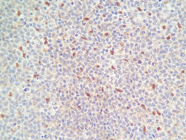
Immunohistochemistry-Paraffin: PD-L1 Antibody [NBP1-76769] - B7-H1/PD-L1/CD274 Antibody [NBP1-76769] - Human tonsil tissue with PD-L1 antibody at 5 ug/mL.
Immunohistochemistry-Paraffin: PD-L1 Antibody [NBP1-76769] - Staining of human heart tissue with antibody at 2.5 ug/mL.
Immunohistochemistry-Paraffin: PD-L1 Antibody [NBP1-76769] - PD-L1/B7-H1 Antibody [NBP1-76769] - PD-L1 in rat heart tissue with at 5 ug/mL.
Flow Cytometry: PD-L1 Antibody [NBP1-76769] - B7-H1/PD-L1/CD274 Antibody [NBP1-76769] - Analysis of A-20 cells using B7-H1/PD-L1/CD274 antibody at 0.5 ug/mL. Green: Isotype control. Red : B7-H1/PD-L1/CD274 antibody.
Immunofluorescence: PD-L1 Antibody [NBP1-76769] - Human Heart cells with PD-L1 antibody at 20 ug/mL.
Dual RNAscope ISH-IHC: PD-L1 Antibody [NBP1-76769] - PDCD1 mRNA (red) and PD-L1 protein (green) were detected in formalin-fixed paraffin-embedded tissue sections of human lung cancer. ACD's Integrated Co-Detection ...read more
Abrogation of autophagy by siRNAs targeting autophagy-related genes in gastric cancer cells induced tumor-intrinsic PD-L1 expression. a Inhibition of autophagy by knockdown of ATG5 in AGS & NCI-n87 gastric cancer cells ...read more
Effects of autophagy inhibitors in combination with IFN-gamma on expression of PD-L1 in gastric cancer cell lines. a The effect of chloroquine (CQ) or 3-MA on expression of PD-L1 with or without INF-gamma for 24 h ...read more
Western Blot: PD-L1 Antibody - BSA Free [NBP1-76769] - ROS mediated PD-L1 upregulation in FFA-treated LO2 cells. (a) Intracellular ROS in LO2 cells before & after FFA treatment measured by DCFH-DA. (b–d) Expression ...read more
Western Blot: PD-L1 Antibody - BSA Free [NBP1-76769] - Inhibition of NF-kappa B or STAT3 downregulated PD-L1 expression in adi-CM model. a PD-L1 protein levels in HepG2 cells treated with different concentration of ...read more
Western Blot: PD-L1 Antibody - BSA Free [NBP1-76769] - PD-L1 expression levels in liver samples & FFA-treated LO2 cells. (a–d) PD-L1 expression & CD8+ T cells in the liver tissues determined by immunohistochemistry. ...read more
Western Blot: PD-L1 Antibody - BSA Free [NBP1-76769] - Inhibition of NF-kappa B or STAT3 downregulated PD-L1 expression in adi-CM model. a PD-L1 protein levels in HepG2 cells treated with different concentration of ...read more
Western Blot: PD-L1 Antibody - BSA Free [NBP1-76769] - ROS/ZNF24/PD-L1 pathway activation in NAFLD models. (a) Intracellular ROS in hepatocytes in NAFLD significantly increased. (b) JC-1 probes were used to detect ...read more
Western Blot: PD-L1 Antibody - BSA Free [NBP1-76769] - PD-L1 limits liver injury in NAFLD models. (a & b) Western blot determined the effect of siRNA against PD-L1. (c) After PD-L1 knockdown, mRNA expression levels of ...read more
Western Blot: PD-L1 Antibody - BSA Free [NBP1-76769] - PD-L1 knockdown aggravated the damage of CD8 + T cells to FFA-treated LO2 cells. (a & b) Western blot determined the effect of siRNA against PD-L1. Coculturing ...read more
Western Blot: PD-L1 Antibody - BSA Free [NBP1-76769] - Upregulated expression of PD‐L1 in cisplatin‐resistant cells through IL‐6/STAT3. The cisplatin resistant CAL27cis & Detroit‐562cis cells had increased ...read more
Western Blot: PD-L1 Antibody - BSA Free [NBP1-76769] - ZNF24 promoted PD-L1 expression through binding to its promoter in FFA-treated LO2 cells. (a) A schematic of the target sites (wild & mutant) of ZNF24 in the ...read more
Western Blot: PD-L1 Antibody - BSA Free [NBP1-76769] - LfcinB reduced IL‐6‐dependent PD‐L1 expression in cisplatin‐resistant HNSCC cells (A‐C) & inhibited the xenograft growth from CAL27cis & Detroit‐562cis ...read more
Western Blot: PD-L1 Antibody - BSA Free [NBP1-76769] - Identification of ZNF24–UBE2I protein interactions. (a & b) Analysis of protein interactions between ZNF24 & UBE2I using the BioGRID database, further confirmed ...read more

Product Details

Summary
Reactivity Hu, Mu, RtSpecies Glossary
Applications WB, ELISA, Flow, ICC/IF, IHC, IP, Dual ISH-IHC, IHC, KD
Clonality
Polyclonal
Host
Rabbit
Conjugate
Unconjugated
Format
BSA Free
Concentration
1 mg/ml

Order Details

Novus Biologicals is part of Bio-Techne

Shop this product on bio-techne.com

PD-L1 Antibody - BSA Free Summary

Immunogen
Antibody was raised against a 17 amino acid synthetic peptide from near the center of human PD-L1. The immunogen is located within amino acids 60-110 of PD-L1.
Specificity
PD-L1 antibody has no cross-reactivity to PD-L2.
Isotype
IgG
Clonality
Polyclonal
Host
Rabbit
Gene
CD274
Purity
Peptide affinity purified
Innovator's Reward
Test in a species/application not listed above to receive a full credit towards a future purchase.

Applications/Dilutions

Dilutions
  • Dual RNAscope ISH-IHC
  • ELISA 1:100 - 1:2000
  • Flow Cytometry 0.5 ug/ml
  • Immunocytochemistry/ Immunofluorescence 1-5 ug/ml
  • Immunohistochemistry Whole-Mount
  • Immunohistochemistry 1:10-1:500
  • Immunohistochemistry-Frozen
  • Immunohistochemistry-Paraffin 1:10-1:500
  • Immunoprecipitation
  • Knockdown Validated
  • Western Blot 1:1000
Application Notes
Use in Immunohistochemistry Whole-Mount reported in scientific literature (PMID:34944780).Use in ICC/IF reported in scientific literature (PMID:33220359) Use in IHC-Frozen was reported in scientific literature (PMID: 28402953). Use in immunoprecipitation reported in scientific literature (PMID: 28978117)..
Theoretical MW
37 kDa.
Disclaimer note: The observed molecular weight of the protein may vary from the listed predicted molecular weight due to post translational modifications, post translation cleavages, relative charges, and other experimental factors.
Control Peptide
PD-L1 Protein (NBP1-76769PEP)
Reviewed Applications
Read 5 Reviews rated 3.2
using
NBP1-76769 in the following applications:

Publications
Read Publications using
NBP1-76769 in the following applications:

Packaging, Storage & Formulations

Storage
Store at 4C short term. Aliquot and store at -20C long term. Avoid freeze-thaw cycles.
Buffer
PBS
Preservative
0.02% Sodium Azide
Concentration
1 mg/ml
Purity
Peptide affinity purified

Alternate Names for PD-L1 Antibody - BSA Free

  • Avelumab
  • B7-H
  • B7H1
  • B7-H1
  • B7H1PDCD1L1
  • CD274 antigenMGC142294
  • CD274 molecule
  • CD274
  • PDCD1L1
  • PDCD1LG1
  • PDCD1LG1MGC142296
  • PDL1
  • PD-L1
  • PD-L1B7 homolog 1
  • PDL1PDCD1 ligand 1
  • programmed cell death 1 ligand 1
  • Programmed death ligand 1

Background

Programmed-death ligand 1 (PD-L1), also known as CD274 and B7-H1, is a 33 kDa type I glycoprotein containing 290 amino acids (aa) belonging to the protein B7 family and serves as part of an immune checkpoint (1,2). PD-L1 contains an Ig-V and Ig-C-like extracellular domain, a transmembrane domain, and a cytoplasmic tail lacking canonical signaling motifs (2,3). PD-L1 is the ligand that binds the receptor programmed-death 1 (PD-1) which is highly expressed on active T cells (1-3). PD-L1 is typically upregulated by tumor cells and antigen presenting cells (APCs), but also expressed on T cells, B cells, macrophages, dendritic cells (DC), mast cells, and some non-immune cell types (1-3). In addition to the membrane-bound, PD-L1 is released from cells both in soluble form and bound to extracellular vesicles (4).

PD-L1 binding with receptor PD-1 results in phosphorylation of in the inhibitory tyrosine-based switch motif (ITSM) domain of PD-1, which leads to recruitment of Src homology 2 domain-containing protein tyrosine-phosphatase 2 (SHP-2) and eventual downstream phosphorylation of spleen tyrosine kinase (Syk) and phospholipid inositol-3-kinase (PI3K) (1,3). Under normal conditions, the PD-L1/PD-1 signaling axis helps maintain immune tolerance and prevent destructive immune responses by inhibiting T cell activity such as proliferation, survival, cytokine production, and cytotoxic T lymphocyte (CTL) cytotoxicity (1-3). In the tumor microenvironment (TME), however, the PD-L1/PD-1 signaling axis is hijacked to promote tumor cell survival and limit anti-tumor immune response (1,3). More precisely, tumor cells can escape killing and immune surveillance due to T cell exhaustion and apoptosis (1-3).

Given the role the PD-L1/PD-1 signaling axis plays in tumor cells' ability to evade immune surveillance, it has become a target of several immunotherapeutic agents in recent years (3,5). Antibody immunotherapies that target these inhibitory checkpoint molecules has shown great promise for cancer treatment (3,5). PD-L1 and PD-1 blocking agents have been approved for treatment in a number of cancers including melanoma, non-small cell lung cancer (NSCLC), urothelial carcinoma, and Merkel-cell carcinoma (3,5). In many cancers the expression of PD-L1 in the TME has predictive value for response to blocking agents (3). Pembrolizumab, for example, is a PD-1 inhibitor that has been approved by the FDA as a second-line therapy for treatment of metastatic NSCLC in patients whose tumors express PD-L1 with a Tumor Proportion Score (TPS) greater than 1%, but also for first-line treatment in cases where patients' tumors expression PD-L1 with a TPS greater than 50%) (5). The most promising cancer immunotherapy treatments seem to point to combination therapy with both anti-cancer drugs (e.g. Gefitibin, Metformin, Etoposide) with PD-L1/PD-1 antibody blockade inhibitors (e.g. Atezolizumab, Nivolumab) (6).

References

1. Han, Y., Liu, D., & Li, L. (2020). PD-1/PD-L1 pathway: current researches in cancer. American journal of cancer research, 10(3), 727-742.

2. Jiang, Y., Chen, M., Nie, H., & Yuan, Y. (2019). PD-1 and PD-L1 in cancer immunotherapy: clinical implications and future considerations. Human vaccines & immunotherapeutics, 15(5), 1111-1122. https://doi.org/10.1080/21645515.2019.1571892

3. Sun, C., Mezzadra, R., & Schumacher, T. N. (2018). Regulation and Function of the PD-L1 Checkpoint. Immunity, 48(3), 434-452. https://doi.org/10.1016/j.immuni.2018.03.014

4. Cha, J. H., Chan, L. C., Li, C. W., Hsu, J. L., & Hung, M. C. (2019). Mechanisms Controlling PD-L1 Expression in Cancer. Molecular cell, 76(3), 359-370. https://doi.org/10.1016/j.molcel.2019.09.030

5. Tsoukalas, N., Kiakou, M., Tsapakidis, K., Tolia, M., Aravantinou-Fatorou, E., Baxevanos, P., Kyrgias, G., & Theocharis, S. (2019). PD-1 and PD-L1 as immunotherapy targets and biomarkers in non-small cell lung cancer. Journal of B.U.ON. : official journal of the Balkan Union of Oncology, 24(3), 883-888.

6. Gou, Q., Dong, C., Xu, H., Khan, B., Jin, J., Liu, Q., Shi, J., & Hou, Y. (2020). PD-L1 degradation pathway and immunotherapy for cancer. Cell death & disease, 11(11), 955. https://doi.org/10.1038/s41419-020-03140-2

Limitations

This product is for research use only and is not approved for use in humans or in clinical diagnosis. Primary Antibodies are guaranteed for 1 year from date of receipt.

Customers Who Viewed This Item Also Viewed...

AF1086
Species: Hu
Applications: Block, CyTOF-ready, Dual ISH-IHC, ELISA(Cap), ELISA(Det), ELISA(Sta), Flow, IHC, WB
MAB1224
Species: Hu
Applications: IHC, WB
7268-CT
Species: Hu
Applications: BA
MAB140
Species: Hu
Applications: CyTOF-ready, Dual ISH-IHC, ELISA(Cap), ELISA(Det), ELISA(Sta), Flow, IHC, Neut
MAB7158
Species: Hu, Mu
Applications: IHC, WB
NB100-524
Species: Hu, Mu
Applications: Flow-IC, Flow, ICC/IF, IHC, IHC-Fr, IHC-P, IP, WB
NBP2-79843
Species: Hu
Applications: CyTOF-ready, ELISA, Flow, ICC/IF, IHC, IHC-P, PA, WB
DY417
Species: Mu
Applications: ELISA
NBP1-49045
Species: Mu, Rt
Applications: Cell Depl, CyTOF-ready, Flow, ICC/IF, IHC, IHC-Fr, IHC-P, IP, InhibTFunc
NBP1-19371
Species: Ca, Hu, Mu, Po, Rb, Rt
Applications: Dual ISH-IHC, Flow, ICC/IF, IHC, IHC-Fr, IHC-P, Simple Western, WB
202-IL
Species: Hu
Applications: BA
NBP2-25208
Species: Hu, Mu, Rt
Applications: Flow, ICC/IF, IHC, IHC-Fr, IHC-P, KO, WB
NBP1-72042
Species: Hu, Mu, Rt
Applications: ICC/IF, IHC, IHC-P, PEP-ELISA, WB
MAB342
Species: Hu
Applications: AgAct, ICC, WB
NB100-39002
Species: Bv, Hu, Mu, Rt
Applications: Dual ISH-IHC, Flow, IB, ICC/IF, IHC, IHC-P, IP, Single-Cell Western, WB
AF165
Species: Hu
Applications: CyTOF-ready, Flow, WB
6507-IL/CF
Species: Hu
Applications: BA
AF169
Species: Hu
Applications: IHC, WB

Publications for PD-L1 Antibody (NBP1-76769)(40)

We have publications tested in 3 confirmed species: Human, Mouse, Rat.

We have publications tested in 12 applications: B/N, ICC/IF, IF/IHC, IHC-F, IHC-P, IHC-WhMt, IP, Immunohistochemistry, Immunoprecipitation, KD, WB, Western Blot.


Filter By Application
B/N
(1)
ICC/IF
(4)
IF/IHC
(5)
IHC-F
(1)
IHC-P
(3)
IHC-WhMt
(1)
IP
(2)
Immunohistochemistry
(2)
Immunoprecipitation
(1)
KD
(1)
WB
(12)
Western Blot
(2)
All Applications
Filter By Species
Human
(16)
Mouse
(9)
Rat
(2)
All Species
Showing Publications 1 - 10 of 40. Show All 40 Publications.
Publications using NBP1-76769 Applications Species
Zhong Y, Li MY, Han L et al. Galangin inhibits programmed cell death-ligand 1 expression by suppressing STAT3 and MYC and enhances T cell tumor-killing activity Phytomedicine : international journal of phytotherapy and phytopharmacology 2023-07-01 [PMID: 37267692] (WB, Human)

Details:
1:2000 WB dilution
WB Human
Mei‐Chih Chen, Meng‐Yu Hung, Chih‐Ming Pan, Shi‐Wei Huang, Chia‐Ing Jan, Yu‐Hsuan Li, Shao‐Chih Chiu, Der‐Yang Cho Pemetrexed combined with dual immune checkpoint blockade enhances cytotoxic T lymphocytes against lung cancer Cancer Science 2023-04-19 [PMID: 37017116]
Caron JM, Han X, Lary CW et al. Targeting the secreted RGDKGE collagen fragment reduces PD?L1 by a proteasome?dependent mechanism and inhibits tumor growth Oncology reports 2023-02-01 [PMID: 36633146] (ICC/IF, Mouse) ICC/IF Mouse
Dong M, Qian M, Ruan Z CUL3/SPOP complex prevents immune escape and enhances chemotherapy sensitivity of ovarian cancer cells through degradation of PD-L1 protein Journal for immunotherapy of cancer 2022-10-01 [PMID: 36198437] (IP, WB, Human) IP, WB Human
Luo Z, Liao T, Zhang Y et al. Ex vivo anchored PD L1 functionally prevent in vivo renal allograft rejection Bioengineering & Translational Medicine 2022-04-06 [PMID: 36176616] (WB, ICC/IF, IHC-P, Rat) WB, ICC/IF, IHC-P Rat
Kozak K, Szumera-Cieckiewicz A. 1441PPROGRAMMED DEATH LIGAND-1 (PD-L1) EXPRESSION AND PROGNOSTIC VALUE IN SYNOVIAL SARCOMA. Annals of Oncology. 2014-01-01
Riondato F, Colitti B, Rosati S et al. A method to test antibody cross-reactivity toward animal antigens for flow cytometry Cytometry. Part A : the journal of the International Society for Analytical Cytology 2022-09-26 [PMID: 36161760]
Sakuma K, Kii T, Takahashi H et al. An In Vivo Study of Local Administration of Low-dose Anti-PD-1 Antibody Using an Oral Cancer Cell Line Anticancer research 2022-09-01 [PMID: 36039414]
Yang W, Han B, Chen Y, Geng F SAAL1, a novel oncogene, is associated with prognosis and immunotherapy in multiple types of cancer Aging 2022-08-13 [PMID: 35963646] (WB, Human) WB Human
Dong G, Huang X, Chen R et al. Increased PD-L1 Restricts Liver Injury in Nonalcoholic Fatty Liver Disease Oxidative Medicine and Cellular Longevity 2022-05-16 [PMID: 35615575] (Immunohistochemistry, Immunoprecipitation, Western Blot) Immunohistochemistry, Immunoprecipitation, Western Blot
Show All 40 Publications.

Reviews for PD-L1 Antibody (NBP1-76769) (5) 3.25

Average Rating: 3.2
(Based on 5 reviews)
We have 5 reviews tested in 4 species: Human, Mouse, Canine, Human and Mouse.

Reviews using NBP1-76769:
Filter by Applications
IHC-P
(2)
ICC
(1)
WB
(2)
All Applications
Filter by Species
Human
(2)
Mouse
(1)
Canine
(1)
Human and Mouse
(1)
All Species
Images Ratings Applications Species Date Details
Immunohistochemistry-Paraffin PD-L1 NBP1-76769
Enlarge
1
reviewed by:
Valeria Martini
IHC-P Canine 08/27/2020
View

Summary

ApplicationImmunohistochemistry-Paraffin
Sample TestedFFPE
SpeciesCanine
LotB

Comments

CommentsParaffin-embedded tissue blocks of 2 different samples were cut into 2 μm sections.
Automated deparaffinization and rehydration was performed by immersing the slides in Xylene, in graded alcohol series and finally in water as indicated.
0.3% hydrogen peroxide was used to block endogenous peroxidase activity for 30 minutes.
Antigen retrieval was performed for each sample both in citrate 10 mM (pH 6.0) buffer and in Tris-EDTA 10 mM (pH 9.0) buffer at 98°C for 30 minutes.
Primary antibody was used both at final dilution of 5 ug/mL and 10 ug/mL in 1.5% horse serum in PBS, and sections were incubated for 2 hours at room temperature.
Incubation with biotinylated secondary antibody and streptavidin/HRP complex were performed using the VECTASTAIN® Universal Quick Kit Peroxidase, R.T.U. (Vector Laboratories) following manufacturer’s instructions.
DAB was added to tissue sections until brown color developed (3 minutes), then slides were moved to water for 5 minutes and counterstained in hematoxylin.
Finally, slides were dehydrated and mounted.
*Low Rating Note: Novus Innovators Program - new species or application used on a primary antibody.
Immunocytochemistry PD-L1 NBP1-76769
Enlarge
3
reviewed by:
Verified Customer
ICC Mouse 05/12/2018
View

Summary

ApplicationImmunocytochemistry
Sample Testedpancreas cancer cells
SpeciesMouse
LotNBP1-76769

Comments

Commentsdilution 1:100 overnight incubation @ 4 C
Western Blot PD-L1 NBP1-76769
Enlarge
5
reviewed by:
Verified Customer
WB Human 03/02/2017
View

Summary

ApplicationWestern Blot
Sample Testedhuman melanoma
SpeciesHuman
Lot7315-1303
  4
reviewed by:
Verified Customer
IHC-P Human and Mouse 01/18/2017
View

Summary

ApplicationImmunohistochemistry-Paraffin
Sample TestedPancreatic cancer tissue,transgenic mouse pancreas
SpeciesHuman and Mouse
Lot8293-1504-2
Western Blot PD-L1 NBP1-76769
Enlarge
3
reviewed by:
Verified Customer
WB Human 11/09/2016
View

Summary

ApplicationWestern Blot
Sample TestedHCT-116 colon cancer cell line w/wo drug treatment
SpeciesHuman

Comments

CommentsI tested protein lording amount, 25, 30, and 50 ug to optimize the PD-L1 expression.
Blocking buffer was Li-COR synthetic protein blocking buffer for 1 hr.
The dilution of Primary antibody(PD-L1, NBP1-76769) were 1:500, 1:1000 and 1:2000 that I tested.
Secondary antibody, Li-COR, IRdye 800CW goat anti-rabbit, 1:5000 dilution for 45 min.

Product General Protocols

Video Protocols

WB Video Protocol
ICC/IF Video Protocol

FAQs for PD-L1 Antibody (NBP1-76769). (Showing 1 - 1 of 1 FAQs).

  1. What can I use as positive controls for B7-H1 (NBP1-76769) other than the myocardial tissue?
    • For CD274 (B7-H1), this protein is highly expressed in the heart, skeletal muscle, placenta and lung ( uniprot site). It can also be found at lower levels in other tissues as well: protein atlas site.

Secondary Antibodies


Isotype Controls

Additional PD-L1 Products

Research Areas for PD-L1 Antibody (NBP1-76769)

Find related products by research area.

Blogs on PD-L1.

Unlocking the Potential of Biosimilars in Immuno-Oncology
By Jennifer Jones, M.S.Biosimilar Antibodies: Imitation Meets InnovationIn the ever-evolving medical landscape, a new class of pharmaceuticals is emerging as a game-changer, poised to transform the way we approach...  Read full blog post.

Synthetic Biotic Medicine as Immunotherapy Against Cancer: Evidence From Arginine-Producing Engineered Bacteria
By Jamshed Arslan, Pharm D, PhDWhat do nuts, dairy and red meat have in common? In addition to the fact that they are all edible, one of the answers is L-arginine. This amino acid improves T cell’s respons...  Read full blog post.

mFluor Violet Conjugated Antibodies

Customers Who Bought This Also Bought

Contact Information

Product PDFs

Calculators

Concentration Calculator

The concentration calculator allows you to quickly calculate the volume, mass or concentration of your vial. Simply enter your mass, volume, or concentration values for your reagent and the calculator will determine the rest.

=
÷

Recent Reviews

3.2
5
1
4
1
3
2
2
0
1
1

Valeria Martini
08/27/2020
Application: IHC-P
Species: Canine

Verified Customer
05/12/2018
Application: ICC
Species: Mouse

Verified Customer
03/02/2017
Application: WB
Species: Human

Bioinformatics

Gene Symbol CD274
Uniprot