

Submit your event on Plasma Membrane Markers to be featured.
Plasma Membrane MarkersWhat is the Plasma Membrane?The plasma membrane is a selectively permeable lipid bilayer that separates the cytoplasm and intracellular components from the extracellular environment of a cell. It is a highly dynamic structure comprised of lipids and proteins, responsive to extracellular signals, like cytokines, hormones, and ions, resulting in intracellular signaling cascades. Movement of substances across the plasma membrane occurs via passive or active transport, the latter of which requires the expenditure of energy. Smaller molecules can cross the cell membrane by passive diffusion whereas larger, more polar molecules generally require active transporter-mediated entry. The plasma membrane also serves as the attachment point for the intracellular cytoskeleton, which helps to determine cell shape and motility. 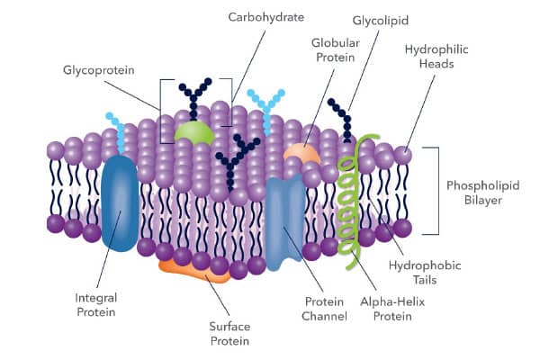
Illustration of protein organization within the lipid bilayer to form the structure of the plasma membrane. The membrane forms a semipermeable barrier between the extracellular and intracellular compartments and provides interaction points with the cytoskeleton. Plasma Membrane Markers and ApplicationsPlasma Membrane LipidsThe phospholipid bilayer consists of two layers of phospholipids with the hydrophilic heads facing the extra- and intracellular environments and hydrophobic tails in the middle. In addition to phospholipids (e.g. phosphatidylserine (PtdSer), phosphatidylcholine (PtdCho)), other lipids, such as sphingolipids (e.g. sphingomyelin (SM)) and cholesterol are abundant in the plasma membrane. Lipids are essential for cell membrane energy storage, compartmentalization, membrane fluidity, and signaling. View Fluorescent Lipid Probes & Cell Membrane Stains from Tocris Plasma Membrane Proteins Common types of plasma membrane proteins include pumps and transporters, enzymes, receptors, and anchors. For example, G-protein coupled receptors (GPCRs) are a large family of receptors that convert extracellular messages into intracellular signaling cascades. The GPCR family of proteins includes important cytokine and chemokine receptors responsible for cell trafficking, like CCR7 and CXCR4. Sodium Potassium ATPase and Pma1 are two common membrane ATPase pumps that have traditionally served as general plasma membrane markers. Plasma membrane proteins also serve as drug candidate targets for the many pathologies associated with dysfunctional membrane proteins. Potential therapeutics can affect the protein function by blocking transport or inhibiting ligand binding, for example, resulting in cellular signaling modifications. Antibodies to markers for the plasma membrane can be used in a wide range of applications including immunocytochemistry (ICC)/immunofluorescence (IF), immunohistochemistry (IHC), western blot, ELISA, flow cytometry, and immunoprecipitation (IP).  and ABCC1 (green) expression, ICC/IF.")
Immunofluorescent image depicting the cellular model of the blood-cerebrospinal fluid (CSF) barrier. Choroidal epithelial cells were stained with Mouse Anti-Sodium Potassium ATPase Alpha 1 Monoclonal Antibody (Catalog #NB300-146) (red) and Rabbit Anti-ABCC1 Polyclonal Antibody (green), highlighting the respective apical and basolateral membrane localization. (Left) Close up image of a single cell showing the polarity of distribution between Na+K+ ATPase and ABCC1, and the nuclei (blue). (Right) Confocal analysis of cells in z-stack. Arrows point to the lateral cellular membranes, whereas the arrowheads emphasize the basal labeling of ABCC1. Image collected and cropped by CiteAb from the following publication (//dx.plos.org/10.1371/journal.pone.0150945), licensed under a CC-BY 4.0 license. Common Plasma Membrane Markers and Function:
Note: The markers listed in the table is not an exhaustive list of all plasma membrane markers. View Plasma Membrane Marker Reagents
Plasma Membrane Damage, Repair, and Role in Disease PathologyPlasma Membrane Damage and RepairInsults to the plasma membrane can result from a variety of different causes including mechanical, chemical, microbial, immune, or intracellular mechanisms. A physical breach or chemical disruption can induce plasma membrane damage and leave the cell susceptible to further injury. Chemical damage through radiation or reactive oxygen species (ROS) can make mechanical stress like nanoruptures and pore formation more likely. Many cell death pathways such as apoptosis, necroptosis, and ferroptisis are characterized, in part, by plasma membrane damage. One of the earliest signs of apoptosis is transition of PtdSer from the inner membrane to the outer membrane. Novus Biologicals’ Polarity Sensitive Indicator of Viability and Apoptosis (pSIVA) contains an Annexin-based probe to detect apoptosis by PtdSer externalization, both irreversible and transient. Our pSIVA Apoptosis Detection Kits are suitable for multiple applications including flow cytometry and ICC/IF. 
Flow Cytometry analysis showing either untreated or Staurosporine treated Jurkat cells stained with Polarity Sensitive Indicator of Viability Apoptosis (pSIVA) Kit [IANBD] (Catalog #NBP2-29611). Results show increasing positive population staining percentage with the pSIVA Kit in the Staurosporine-treated cells, indicative of cell death. The plasma membrane follows four stages of repair after a physical breach:
The Role of the Plasma Membrane in Disease PathogenesisDefective plasma membrane repair pathway and the inability to restore plasma membrane integrity can cause or exacerbate the pathogenesis of a variety of diseases such as inflammatory bowel disease, neurodegenerative disorders, and muscular dystrophies (MDs). Muscular dystrophies, for example, are often characterized by increased membrane permeability, resulting in degradation of the muscle and loss of function. Similarly, plasma membrane integrity is lost during Alzheimer’s disease and Parkinson’s disease pathogenesis, as the disease-associated proteins form aggregates. ReferencesAmmendolia, D. A., Bement, W. M., & Brumell, J. H. (2021). Plasma membrane integrity: implications for health and disease. BMC biology, 19(1), 71. https://doi.org/10.1186/s12915-021-00972-y Andrews, N. W., & Corrotte, M. (2018). Plasma membrane repair. Current biology : CB, 28(8), R392–R397. https://doi.org/10.1016/j.cub.2017.12.034 Bernardino de la Serna, J., Schütz, G. J., Eggeling, C., & Cebecauer, M. (2016). There Is No Simple Model of the Plasma Membrane Organization. Frontiers in cell and developmental biology, 4, 106. https://doi.org/10.3389/fcell.2016.00106 Burns, K. E., & Delehanty, J. B. (2017). Targeting therapeutics to the plasma membrane: opportunities for nanoparticle-mediated delivery abound. Therapeutic delivery, 8(5), 235–237. https://doi.org/10.4155/tde-2016-0091 Casares, D., Escribá, P. V., & Rosselló, C. A. (2019). Membrane Lipid Composition: Effect on Membrane and Organelle Structure, Function and Compartmentalization and Therapeutic Avenues. International journal of molecular sciences, 20(9), 2167. https://doi.org/10.3390/ijms20092167 Dias, C., & Nylandsted, J. (2021). Plasma membrane integrity in health and disease: significance and therapeutic potential. Cell discovery, 7(1), 4. https://doi.org/10.1038/s41421-020-00233-2 Kraft M. L. (2013). Plasma membrane organization and function: moving past lipid rafts. Molecular biology of the cell, 24(18), 2765–2768. https://doi.org/10.1091/mbc.E13-03-0165 Lukiw W. J. (2013). Alzheimer's disease (AD) as a disorder of the plasma membrane. Frontiers in physiology, 4, 24. https://doi.org/10.3389/fphys.2013.00024 Parton, R. G., Tillu, V. A., & Collins, B. M. (2018). Caveolae. Current biology : CB, 28(8), R402–R405. https://doi.org/10.1016/j.cub.2017.11.075 Van Campenhout, R., Muyldermans, S., Vinken, M., Devoogdt, N., & De Groof, T. (2021). Therapeutic Nanobodies Targeting Cell Plasma Membrane Transport Proteins: A High-Risk/High-Gain Endeavor. Biomolecules, 11(1), 63. https://doi.org/10.3390/biom11010063 Yang, N. J., & Hinner, M. J. (2015). Getting across the cell membrane: an overview for small molecules, peptides, and proteins. Methods in molecular biology (Clifton, N.J.), 1266, 29–53. https://doi.org/10.1007/978-1-4939-2272-7_3
|


 and probed with Rabbit Anti-Glut1 Antibody, WB.")