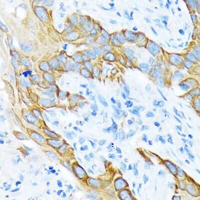
Novus Biologicals products are now on bio-techne.com

Cytokeratin, pan Antibody

Images

 
Immunohistochemistry-Paraffin: Cytokeratin, pan Antibody [NB600-579] - Human skin stained with Cytokeratin antibody.

Product Details

Summary
Reactivity Hu, Mu, Bv, PmSpecies Glossary
Applications IHC
Clonality
Polyclonal
Host
Rabbit
Conjugate
Unconjugated

Order Details

Cytokeratin, pan Antibody Summary

Immunogen
This Cytokeratin, pan Antibody was developed against pan Cytokeratin isolated from bovine muzzle epidermis.
Localization
Cytoplasmic
Marker
Epithelial marker
Specificity
NB600-579 has cross-reactivity with cytokeratins of 58, 56, 52, 60, 51, 48 and 68 kDa molecular weight. The antibody is well suited for the staining of a broad spectrum of human keratins.
Isotype
IgG
Clonality
Polyclonal
Host
Rabbit
Gene
KRT1
Purity
Protein A purified
Innovator's Reward
Test in a species/application not listed above to receive a full credit towards a future purchase.

Applications/Dilutions

Dilutions
  • Immunohistochemistry 1:50-1:100
  • Immunohistochemistry-Frozen 1:75 - 1:200
  • Immunohistochemistry-Paraffin 1:50-1:100
Application Notes
IHC-P: recommended pretreatment of HistoZyme. Recommended incubation time of 30 min at RT.
Control
Human Skin Whole Tissue Lysate (Adult Whole Normal)
Publications
Read Publications using
NB600-579 in the following applications:

Reactivity Notes

Monkey reactivity reported in scientific literature (PMID: 28916434). Mouse reactivity reported in scientific literature (PMID: 32421534).

Packaging, Storage & Formulations

Storage
Store at 4C. Do not freeze.
Buffer
PBS (pH 7.4), 0.2% BSA, Tween-20
Preservative
0.05% Sodium Azide
Purity
Protein A purified

Alternate Names for Cytokeratin, pan Antibody

  • AEI2
  • AEI2;CK1;EHK;EHK1;EPPK;K1;Keratin, type II cytoskeletal 1;KRT1A;NEPPK
  • CK1
  • Cytokeratin, pan
  • EHK
  • EPPK
  • K1
  • KRT1A
  • NEPPK

Background

Cytokeratins are a family of intermediate filamentous proteins that are expressed by epithelial cells (1,2). Cytokeratins range in size with a theoretic molecular weight varying from approximately 40 kDa to 68 kDa (2,3). The cytokeratin family consists of 20 polypeptides that are further divided into two main groups based on isoelectric point and molecular weight (1-3). The type I group are smaller, acidic polypeptides designated as cytokeratin 9 through cytokeratin 20 (CK9 - CK20) (1-4). Conversely, CK1 - CK8 belong to the type II group, classified as larger, basic or neutral polypeptides (1-4). Structurally, cytokeratins have homologous basic structure with other intermediate filaments; they possess a 300-315 amino acid (aa) central helical region that consists of four conserved domains (1A, 2A, 1B, and 2B) which are separated by linker domains (L1, L12, and L2) (1,5). Additionally, flanking this central region, both the amino- and carboxyl-terminal ends have a homologous subdomain (H), a variable domain (V), and charged end subdomains (E) (1). Furthermore, the central rod of one cytokeratin monomer binds with another monomer to form a coiled-coil dimer which subsequently binds another dimer to form a tertramer (3). Finally, many tetramers join together to ultimately form an intermediate filament of approximately 10nm in diameter (1-3, 5). Cytokeratins are expressed as pairs, typically with a type I and type II member; for example, CK10 pairs with CK1 (1,3).

Epithelial cells express multiple subtypes of cytokeratins which can be used to classify epithelial cell type or differentiation status, as well tumor progression or diagnosis (2). Cytokeratins are important for both stability and integrity of epithelial cells and function in intracellular signaling, from wound healing to apoptosis (1). Cytokeratins are useful immunohistochemistry tumor markers and antibodies to cytokeratins are a common pathological tool (1,3,6). Cytokeratin pan antibody is an antibody cocktail mixture that can detect multiple cytokeratins and reacts to multiple epithelial tissues (1,3,6). For example, AE-1/AE-3 is a commonly used specific pan cytokeratin that detects cytokeratins 1-8, 10, 14-16 and 19 (1,3,6).

Given the role of cytokeratins in the structural integrity of epithelial cells, mutations in cytokeratins have been shown to play a role in a variety of human diseases including epidermolysis bullosa simplex (EBS) (4,5). EBS is an autosomal dominant disorder that is caused by missense mutations in either CK5 or CK14 (5). Other known cytokeratin-related disorders include bullous ichthyosis, a skin disorder characterized by redness, blistering, and hyperkeratosis, and epidermolytic palmoplantar keratoderma (EPPK), which results in hyperkeratosis on the palms and soles of the body (7).

References

1. Awasthi, P., Thahriani, A., Bhattacharya, A., Awasthi, P., & Keratins, B. A. (2016). Keratins or cytokeratins: a review article. Journal of Advanced Medical and Dental Sciences Research. https://10.21276/jamdsr.2016.4.4.30

2. Southgate, J., Harnden, P., & Trejdosiewicz, L. K. (1999). Cytokeratin expression patterns in normal and malignant urothelium: a review of the biological and diagnostic implications. Histology and histopathology. https://doi.org/10.14670/HH-14.657

3. Belaldavar, C., Mane, D. R., Hallikerimath, S., Kale, A. D. (2016). Cytokeratins: Its role and expression profile in oral health and disease. Journal of Oral and Maxillofacial Surgery, Medicine, and Pathology. https://doi.org/10.1016/j.ajoms.2015.08.001.

4. Linder S. (2007). Cytokeratin markers come of age. Tumour biology : the journal of the International Society for Oncodevelopmental Biology and Medicine. https://doi.org/10.1159/000107582

5. Jacob, J. T., Coulombe, P. A., Kwan, R., & Omary, M. B. (2018). Types I and II Keratin Intermediate Filaments. Cold Spring Harbor perspectives in biology. https://doi.org/10.1101/cshperspect.a018275

6. Ordonez N. G. (2013). Broad-spectrum immunohistochemical epithelial markers: a review. Human pathology. https://doi.org/10.1016/j.humpath.2012.11.016

7. McLean, W. H., & Moore, C. B. (2011). Keratin disorders: from gene to therapy. Human molecular genetics. https://doi.org/10.1093/hmg/ddr379

Limitations

This product is for research use only and is not approved for use in humans or in clinical diagnosis. Primary Antibodies are guaranteed for 1 year from date of receipt.

Customers Who Viewed This Item Also Viewed...

NBP2-61736
Species: Hu, Pm, Mu, Rt
Applications: ELISA, Flow, IHC, IHC-P, WB
NBP2-34270
Species: Hu, Mu, Rt
Applications: Flow, ICC/IF, IHC, IHC-Fr, IHC-P, Simple Western
NBP2-61931
Species: Hu, Rt
Applications: Flow, ICC/IF, IHC, IHC-P, WB
NB100-2727
Species: Ca, Hu, Po, Pm
Applications: CyTOF-ready, Flow, ICC/IF, IHC, IHC-P, IP, WB
NBP1-87528
Species: Hu
Applications: ICC/IF, IHC, IHC-P
DNST0
Species: Hu
Applications: ELISA
NBP1-84732
Species: Hu, Mu, Rt
Applications: IHC, IHC-P, WB
NBP2-79718
Species: Hu
Applications: ICC/IF, IHC, IHC-P, PA, WB
NBP3-15243
Species: Hu, Mu, Rt
Applications: ICC/IF, IHC, IHC-P, WB
NBP3-15261
Species: Hu, Mu, Rt
Applications: ICC/IF, IHC, IHC-P, WB
NBP2-67559
Species: Hu
Applications: Flow, ICC/IF, IHC, IHC-P, WB
NBP1-85630
Species: Hu
Applications: IHC, IHC-P, WB
8184-CK
Species: Hu
Applications: EnzAct
NB100-687
Species: Hu, Mu, Rt
Applications: Flow, ICC/IF, IHC, IHC-P, Simple Western, WB
MAB1455
Species: Hu
Applications: CyTOF-ready, Dual ISH-IHC, ICC, IHC, ICFlow, Simple Western, WB
NBP2-16094
Species: Hu, Mu, Ze
Applications: ICC/IF, IHC-WhMt, IHC, IHC-P, KO, WB
NBP2-85137
Species: Hu, Pm, Mu
Applications: IHC, IHC-P, WB
NBP2-12900
Species: Hu, Pm, Mu, Rt
Applications: ICC/IF, IHC, IHC-P, MiAr, Simple Western, WB

Publications for Cytokeratin, pan Antibody (NB600-579)(11)

We have publications tested in 3 confirmed species: Human, Mouse, Monkey.

We have publications tested in 2 applications: IF/IHC, IHC-P.


Filter By Application
IF/IHC
(3)
IHC-P
(3)
All Applications
Filter By Species
Human
(2)
Mouse
(4)
Monkey
(1)
All Species
Showing Publications 1 - 10 of 11. Show All 11 Publications.
Publications using NB600-579 Applications Species
Pietrobono S, Bertolini M, De Vita V et al. CCL3 predicts exceptional response to TGF beta inhibition in basal-like pancreatic cancer enriched in LIF-producing macrophages NPJ Precision Oncology 2024-10-30 [PMID: 39478186]
Felix R De Bie, Yannick Regin, Antoine Dubois, Marianna Scuglia, Tomohiro Arai, Ewout Muylle, David Basurto, Marius Regin, Siska Croubels, Marc Cherlet, Emily A Partridge, Karel Allegaert, Francesca M Russo, Jan A Deprest Prenatal treprostinil improves pulmonary arteriolar hypermuscularization in the rabbit model of congenital diaphragmatic hernia. Biomedicine & pharmacotherapy = Biomedecine & pharmacotherapie 2023-12-11 [PMID: 38086148]
Napso T, Lean SC, Lu M et al. Diet-induced maternal obesity impacts feto-placental growth and induces sex-specific alterations in placental morphology, mitochondrial bioenergetics, dynamics, lipid metabolism and oxidative stress in mice Acta physiologica (Oxford, England) 2022-02-03 [PMID: 35114078] (IF/IHC, Mouse) IF/IHC Mouse
Ionel Sandovici, Aikaterini Georgopoulou, Vicente Pérez-García, Antonia Hufnagel, Jorge López-Tello, Brian Y.H. Lam et al. The imprinted Igf2-Igf2r axis is critical for matching placental microvasculature expansion to fetal growth Developmental Cell 2022-01-10 [PMID: 34963058]
Eber M, Park S, Contino K et al. Osteoblasts derived from mouse mandible enhance tumor growth of prostate cancer more than osteoblasts derived from long bone Journal of Bone Oncology 2021-02-01 [PMID: 33425674] (IHC-P, Mouse) IHC-P Mouse
Xie J, Yuan S, Peng L et al. Antitumor immunity targeting fibroblast activation protein‑ alpha in a mouse Lewis lung carcinoma model Oncol Lett 2020-05-18 [PMID: 32566014]
De Clercq K, Lopez-Tello J, Vriens J, Sferruzzi-Perri A Double-label immunohistochemistry to assess labyrinth structure of the mouse placenta with stereology Placenta 2020-05-01 [PMID: 32421534] (IHC-P, Mouse) IHC-P Mouse
Georges LMC, De Wever O, Galvan JA et al. Cell Line Derived Xenograft Mouse Models Are a Suitable in vivo Model for Studying Tumor Budding in Colorectal Cancer Front Med (Lausanne) 2019-06-27 [PMID: 31316988] (IF/IHC, Human) IF/IHC Human
Slivka, PF;Hsieh, CL;Lipovsky, A;Pratt, SD;Locklear, J;Namovic, MT;McDonald, HA;Wetter, J;Edelmayer, R;Hu, M;Murphy, E;Domanus, M;Lu, C;Duggan, R;King, J;Scott, VE;Donnelly-Roberts, D;Slavin, A;Gopalakrishnan, S;Chung, N;Goedken, ER; Small Molecule and Pooled CRISPR Screens Investigating IL17 Signaling Identify BRD2 as a Novel Contributor to Keratinocyte Inflammatory Responses ACS Chem. Biol. 2019-04-15 [PMID: 30938974] (IHC-P, Human) IHC-P Human
Nicholson SM, Carlesso G, Cheng LI et al. Effects of ICOS+ T cell depletion via afucosylated monoclonal antibody MEDI-570 on pregnant cynomolgus monkeys and the developing offspring Reprod. Toxicol. 2017-09-13 [PMID: 28916434] (Monkey) Monkey
Show All 11 Publications.

Reviews for Cytokeratin, pan Antibody (NB600-579) (0)

There are no reviews for Cytokeratin, pan Antibody (NB600-579). By submitting a review you will receive an Amazon e-Gift Card or Novus Product Discount.
  • Review with no image -- $10/€7/£6/$10 CAD/¥70 Yuan/¥1110 Yen
  • Review with an image -- $25/€18/£15/$25 CAD/¥150 Yuan/¥2500 Yen

Product General Protocols

Find general support by application which include: protocols, troubleshooting, illustrated assays, videos and webinars.

FAQs for Cytokeratin, pan Antibody (NB600-579). (Showing 1 - 1 of 1 FAQs).

  1. Do you have any experience staining lymphoepithelium or epithelial cells in general with this antibody? Is this antibody also working on frozen sections of mice specimens?
    • NB600-579 has been validated for use in IHC on frozen and paraffin-embedded tissues in human, mouse and bovine samples. It should be suitable for your experiments.

Control Lysate(s)

Secondary Antibodies


Isotype Controls

Additional Cytokeratin, pan Products

Array NB600-579

Research Areas for Cytokeratin, pan Antibody (NB600-579)

Find related products by research area.

Blogs on Cytokeratin, pan

There are no specific blogs for Cytokeratin, pan, but you can read our latest blog posts.
Read our latest blog and use the new citation tool on bio-techne.com

Customers Who Bought This Also Bought

Contact Information

Product PDFs

Calculators

Concentration Calculator

The concentration calculator allows you to quickly calculate the volume, mass or concentration of your vial. Simply enter your mass, volume, or concentration values for your reagent and the calculator will determine the rest.

=
÷

Review this Product

Be the first to review our Cytokeratin, pan Antibody and receive a gift card or discount.

Bioinformatics

Gene Symbol KRT1