
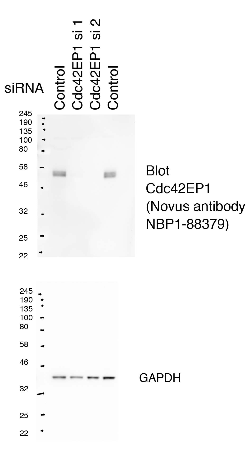
![Western Blot: CDC42EP1 Antibody [NBP1-88379] - HeLa whole cell lysate. Lanes 1 and 4: HeLa cells transfected with control non-targeting siRNA. Lanes 2 and 3: HeLa cells transfected with two different siRNA-targeting Cdc42EP1. Blot probed with cdc42ep1 antibody and reprobed with GAPDH as loading control. Image from verified customer review.](http://images.novusbio.com/fullsize/CDC42EP1-Antibody-Western-Blot-NBP1-88379-img0005.jpg "Western Blot: CDC42EP1 Antibody [NBP1-88379] - HeLa whole cell lysate. Lanes 1 and 4: HeLa cells transfected with control non-targeting siRNA. Lanes 2 and 3: HeLa cells transfected with two different siRNA-targeting Cdc42EP1. Blot probed with cdc42ep1 antibody and reprobed with GAPDH as loading control. Image from verified customer review.")
| Reactivity | HuSpecies Glossary |
| Applications | WB, ICC/IF, IHC, IP, KD |
| Clonality | Polyclonal |
| Host | Rabbit |
| Conjugate | Unconjugated |
| Immunogen | This antibody was developed against Recombinant Protein corresponding to amino acids: SGFCTISRLPRSEKPHDRDRDGSFPSEPGLRRSDSLLSFRLDLDLGPSLLSELLGVMSLPEAPAAETPAPAANPPAPTANPTGPAANPPATTANPPAPAANPSAPAATPTGPAANPPAPAASSTPHGHCPNGV |
| Isotype | IgG |
| Clonality | Polyclonal |
| Host | Rabbit |
| Gene | CDC42EP1 |
| Purity | Immunogen affinity purified |
| Innovator's Reward | Test in a species/application not listed above to receive a full credit towards a future purchase. |
| Dilutions |
|
||
| Application Notes | For IHC-Paraffin, HIER pH 6 antigen retrieval is recommended. ICC/IF fixation Permeabilization, Use PFA/Triton X-100. |
||
| Control Peptide |
|
||
| Reviewed Applications |
|
| Storage | Store at 4C short term. Aliquot and store at -20C long term. Avoid freeze-thaw cycles. |
| Buffer | PBS (pH 7.2) and 40% Glycerol |
| Preservative | 0.02% Sodium Azide |
| Purity | Immunogen affinity purified |


![Western Blot AKT [p Ser473] Antibody [Unconjugated] - Pan Specific](https://images.novusbio.com/images2/Akt3_AF887_Western_Blot_5930.jpg)
![Simple Western AKT [p Ser473] Antibody [Unconjugated] - Pan Specific](https://images.novusbio.com/images2/16332.jpg)
![Intracellular Staining by Flow Cytometry AKT [p Ser473] Antibody [Unconjugated] - Pan Specific](https://images.novusbio.com/images2/Akt3_AF887_Flow_Cytometry_8283.jpg)


| Images | Ratings | Applications | Species | Date | Details | ||||||||||
|---|---|---|---|---|---|---|---|---|---|---|---|---|---|---|---|

Enlarge |
reviewed by:
Stephen Terry |
ICC | Human | 10/25/2018 |
Summary
Comments
|
||||||||||

Enlarge |
reviewed by:
Stephen Terry |
IP | Human | 08/20/2018 |
Summary
Comments
|
||||||||||
Enlarge |
reviewed by:
Stephen Terry |
WB | Human | 08/03/2018 |
Summary
Comments
|
Secondary Antibodies |
Isotype Controls |
Research Areas for CDC42EP1 Antibody (NBP1-88379)Find related products by research area.
|

The concentration calculator allows you to quickly calculate the volume, mass or concentration of your vial. Simply enter your mass, volume, or concentration values for your reagent and the calculator will determine the rest.
5 | |
4 | |
3 | |
2 | |
1 |
| Stephen Terry 10/25/2018 |
||
| Application: | ICC | |
| Species: | Human |
| Stephen Terry 08/20/2018 |
||
| Application: | IP | |
| Species: | Human |
| Stephen Terry 08/03/2018 |
||
| Application: | WB | |
| Species: | Human |
| Gene Symbol | CDC42EP1 |


![Immunocytochemistry/Immunofluorescence: CDC42EP1 Antibody [NBP1-88379] - Confocal Imunofluorescent images of HeLa cells fixed with PFA and stained with Cdc42EP1 antibody at 1:200 (magenta), and phalloidin-Alexa 488 (green) to visualize F-actin. Antibody stains sub nuclear filaments with some co-loclization to actin filaments. Image from verified customer review.](http://images.novusbio.com/fullsize/CDC42EP1-Antibody-Immunocytochemistry-Immunofluorescence-NBP1-88379-img0007.jpg "Immunocytochemistry/Immunofluorescence: CDC42EP1 Antibody [NBP1-88379] - Confocal Imunofluorescent images of HeLa cells fixed with PFA and stained with Cdc42EP1 antibody at 1:200 (magenta), and phalloidin-Alexa 488 (green) to visualize F-actin. Antibody stains sub nuclear filaments with some co-loclization to actin filaments. Image from verified customer review.")
![Immunohistochemistry-Paraffin: CDC42EP1 Antibody [NBP1-88379] - Staining of human stomach shows strong cytoplasmic positivity in glandular cells.](http://images.novusbio.com/fullsize/CDC42EP1-Antibody-Immunohistochemistry-Paraffin-NBP1-88379-img0002.jpg "Immunohistochemistry-Paraffin: CDC42EP1 Antibody [NBP1-88379] - Staining of human stomach shows strong cytoplasmic positivity in glandular cells.")
![Immunocytochemistry/Immunofluorescence: CDC42EP1 Antibody [NBP1-88379] - Staining of human cell line A-431 shows localization to plasma membrane & actin filaments. Antibody staining is shown in green.](http://images.novusbio.com/fullsize/CDC42EP1-Antibody-Immunocytochemistry-Immunofluorescence-NBP1-88379-img0004.jpg "Immunocytochemistry/Immunofluorescence: CDC42EP1 Antibody [NBP1-88379] - Staining of human cell line A-431 shows localization to plasma membrane & actin filaments. Antibody staining is shown in green.")
![Immunoprecipitation: CDC42EP1 Antibody [NBP1-88379] - Lanes 1 and 5: Marker. Lanes 2-4 HeLa cell input controls. Lane 6: IP using 2 mg of Control Rabbit IgG cell lysate. Lane 8: IP using 2 mg Cdc42EP1 cell lysate and Betheyl antibody A302-381A. Lane 10: IP using 2 mg Cdc42EP1 cell lysate and Novus antibody NBP1-88379. Image from verified customer review.](http://images.novusbio.com/fullsize/CDC42EP1-Antibody-Immunoprecipitation-NBP1-88379-img0006.jpg "Immunoprecipitation: CDC42EP1 Antibody [NBP1-88379] - Lanes 1 and 5: Marker. Lanes 2-4 HeLa cell input controls. Lane 6: IP using 2 mg of Control Rabbit IgG cell lysate. Lane 8: IP using 2 mg Cdc42EP1 cell lysate and Betheyl antibody A302-381A. Lane 10: IP using 2 mg Cdc42EP1 cell lysate and Novus antibody NBP1-88379. Image from verified customer review.")


![Immunohistochemistry FGFR3 Antibody (136312) [Unconjugated] - (IIIc)](https://images.novusbio.com/images2/FGF_R3_MAB7662_Immunohistochemistry_23319.jpg)
![Flow Cytometry FGFR3 Antibody (136312) [Unconjugated] - (IIIc)](https://images.novusbio.com/images2/mab7662_human-fgf-r3-iiic-mab-clone-136312-flow-cytometry-2112202112199.jpg)
![Immunocytochemistry FGFR3 Antibody (136312) [Unconjugated] - (IIIc)](https://images.novusbio.com/images2/mab7662_human-fgf-r3-iiic-mab-clone-136312-immunocytochemistry-186202172236.jpg)
 using a 1:1000 dilution of HRP-conjugated Anti-Rabbit IgG Secondary Antibody (Catalog # HAF008). This experiment was conducted under reducing conditions and using Immunoblot Buffer Group 1.")
![Western Blot: Goat anti-Rabbit IgG (H+L) Secondary Antibody [HRP] [NB7160] - Western blot showing vemurafenib treatment in BRAFV600E CRC cells inhibits fission mediator DRP1 with no significant effect on fusion proteins (Mfn1 & 2) using MFN-1 antibody (NBP1-51841) and corresponding secondary antibody, goat anti-rabbit IgG-HRP (NB7160). Image collected and cropped by CiteAb from the following publication (https://pubmed.ncbi.nlm.nih.gov/33738242).](https://images.novusbio.com/images/Goat-anti-Rabbit-IgG-H+L-Secondary-Antibody-HRP-Western-Blot-NB7160-img0001.jpg "Western Blot: Goat anti-Rabbit IgG (H+L) Secondary Antibody [HRP] [NB7160] - Western blot showing vemurafenib treatment in BRAFV600E CRC cells inhibits fission mediator DRP1 with no significant effect on fusion proteins (Mfn1 & 2) using MFN-1 antibody (NBP1-51841) and corresponding secondary antibody, goat anti-rabbit IgG-HRP (NB7160). Image collected and cropped by CiteAb from the following publication (https://pubmed.ncbi.nlm.nih.gov/33738242).")
![Flow Cytometry: Rabbit IgG Isotype Control [NBP2-24891] - Intracellular FACS analysis of mouse TLR6 polyclonal antibody (red), rabbit isotype control (green), RAW cells alone (shaded). Two micrograms of antibodies were used. Goat anti-rabbit FITC (Novus, 20302) was used as secondary.](https://images.novusbio.com/images/Rabbit--Mouse-IgG-Isotype-Control-Flow-Cytometry-NBP2-24891-img0001.jpg "Flow Cytometry: Rabbit IgG Isotype Control [NBP2-24891] - Intracellular FACS analysis of mouse TLR6 polyclonal antibody (red), rabbit isotype control (green), RAW cells alone (shaded). Two micrograms of antibodies were used. Goat anti-rabbit FITC (Novus, 20302) was used as secondary.")


