
![Immunohistochemistry-Paraffin: ATP6V0A1 Antibody [NBP1-89342] - Analysis in human cerebral cortex and tonsil tissues. Corresponding ATP6V0A1 RNA-seq data are presented for the same tissues.](http://images.novusbio.com/fullsize/ATP6V0A1-Antibody-Immunohistochemistry-Paraffin-NBP1-89342-img0015.jpg "Immunohistochemistry-Paraffin: ATP6V0A1 Antibody [NBP1-89342] - Analysis in human cerebral cortex and tonsil tissues. Corresponding ATP6V0A1 RNA-seq data are presented for the same tissues.")
| Reactivity | Hu, Mu, RtSpecies Glossary |
| Applications | WB, ICC/IF, IHC, IP |
| Clonality | Polyclonal |
| Host | Rabbit |
| Conjugate | Unconjugated |
| Immunogen | This antibody was developed against Recombinant Protein corresponding to amino acids: VQFRDLNPDVNVFQRKFVNEVRRCEEMDRKLRFVEKEIRKANIPIMDTGENPEVPFPRDMIDLEANFEKIENELKEINTNQEALKRNFLELTELK |
| Predicted Species | Rat (100%). Backed by our 100% Guarantee. |
| Isotype | IgG |
| Clonality | Polyclonal |
| Host | Rabbit |
| Gene | ATP6V0A1 |
| Purity | Immunogen affinity purified |
| Innovator's Reward | Test in a species/application not listed above to receive a full credit towards a future purchase. |
| Dilutions |
|
||
| Application Notes | For IHC-Paraffin, HIER pH 6 retrieval is recommended. ICC/IF Fixation Permeabilization: Use PFA/Triton X-100. |
||
| Control Peptide |
|
||
| Reviewed Applications |
|
||
| Publications |
|
| Storage | Store at 4C short term. Aliquot and store at -20C long term. Avoid freeze-thaw cycles. |
| Buffer | PBS (pH 7.2), 40% Glycerol |
| Preservative | 0.02% Sodium Azide |
| Purity | Immunogen affinity purified |


| Images | Ratings | Applications | Species | Date | Details | ||||||||
|---|---|---|---|---|---|---|---|---|---|---|---|---|---|

Enlarge |
reviewed by:
Verified Customer |
ICC | Mouse | 10/13/2020 |
Summary
|
Secondary Antibodies |
Isotype Controls |

The concentration calculator allows you to quickly calculate the volume, mass or concentration of your vial. Simply enter your mass, volume, or concentration values for your reagent and the calculator will determine the rest.
| Gene Symbol | ATP6V0A1 |


![Western Blot: ATP6V0A1 Antibody [NBP1-89342] - In Cln1-/- mice V0a1 is misrouted to plasma membrane preventing its interaction with AP-3. Pull-down assay with AP-3 antibody using total lysates from untreated (lane 1) and bromopalmitate-treated (lane 2) WT brain slices to detect V0a1 and its densitometric quantitation (n=4, *P<0.05). HEK-293 cells were transfected with WT GFP-V0a1 and GFP-V0a1-Cys25Ser mutant construct, and pull-down experiments were conducted with AP-3 antibody to detect GFP-V0a1,*P<0.05(n=4). Image collected and cropped by CiteAb from the following publication (//www.nature.com/doifinder/10.1038/ncomms14612), licensed under a CC-BY license.](http://images.novusbio.com/fullsize/ATP6V0A1-Antibody-Western-Blot-NBP1-89342-img0018.jpg "Western Blot: ATP6V0A1 Antibody [NBP1-89342] - In Cln1-/- mice V0a1 is misrouted to plasma membrane preventing its interaction with AP-3. Pull-down assay with AP-3 antibody using total lysates from untreated (lane 1) and bromopalmitate-treated (lane 2) WT brain slices to detect V0a1 and its densitometric quantitation (n=4, *P<0.05). HEK-293 cells were transfected with WT GFP-V0a1 and GFP-V0a1-Cys25Ser mutant construct, and pull-down experiments were conducted with AP-3 antibody to detect GFP-V0a1,*P<0.05(n=4). Image collected and cropped by CiteAb from the following publication (//www.nature.com/doifinder/10.1038/ncomms14612), licensed under a CC-BY license.")
![Western Blot: ATP6V0A1 Antibody [NBP1-89342] - In Cln1-/- mice V0a1 is misrouted to plasma membrane preventing its interaction with AP-3. Western blot analysis and densitometric quantitation of V0a1 in isolated plasma membrane fraction from WT and Cln1-/- mouse brain (n=4, *P<0.05). Image collected and cropped by CiteAb from the following publication (//www.nature.com/doifinder/10.1038/ncomms14612), licensed under a CC-BY license.](http://images.novusbio.com/fullsize/ATP6V0A1-Antibody-Western-Blot-NBP1-89342-img0017.jpg "Western Blot: ATP6V0A1 Antibody [NBP1-89342] - In Cln1-/- mice V0a1 is misrouted to plasma membrane preventing its interaction with AP-3. Western blot analysis and densitometric quantitation of V0a1 in isolated plasma membrane fraction from WT and Cln1-/- mouse brain (n=4, *P<0.05). Image collected and cropped by CiteAb from the following publication (//www.nature.com/doifinder/10.1038/ncomms14612), licensed under a CC-BY license.")
![Immunocytochemistry/Immunofluorescence: ATP6V0A1 Antibody [NBP1-89342] - Staining of human cell line A-431 shows localization to the Golgi apparatus & vesicles. Antibody staining is shown in green.](http://images.novusbio.com/fullsize/ATP6V0A1-Antibody-Immunocytochemistry-Immunofluorescence-NBP1-89342-img0006.jpg "Immunocytochemistry/Immunofluorescence: ATP6V0A1 Antibody [NBP1-89342] - Staining of human cell line A-431 shows localization to the Golgi apparatus & vesicles. Antibody staining is shown in green.")
![Immunohistochemistry-Paraffin: ATP6V0A1 Antibody [NBP1-89342] - Staining of human cerebral cortex shows strong cytoplasmic positivity in neuropil.](http://images.novusbio.com/fullsize/ATP6V0A1-Antibody-Immunohistochemistry-Paraffin-NBP1-89342-img0016.jpg "Immunohistochemistry-Paraffin: ATP6V0A1 Antibody [NBP1-89342] - Staining of human cerebral cortex shows strong cytoplasmic positivity in neuropil.")
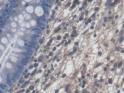
![Immunohistochemistry-Paraffin: ATP6V0A1 Antibody [NBP1-89342] - Staining of human tonsil shows low expression as expected.](http://images.novusbio.com/fullsize/ATP6V0A1-Antibody-Immunohistochemistry-Paraffin-NBP1-89342-img0011.jpg "Immunohistochemistry-Paraffin: ATP6V0A1 Antibody [NBP1-89342] - Staining of human tonsil shows low expression as expected.")
![Immunohistochemistry-Paraffin: ATP6V0A1 Antibody [NBP1-89342] - Staining of human kidney shows strong cytoplasmic postivity in cells in tubules.](http://images.novusbio.com/fullsize/ATP6V0A1-Antibody-Immunohistochemistry-Paraffin-NBP1-89342-img0013.jpg "Immunohistochemistry-Paraffin: ATP6V0A1 Antibody [NBP1-89342] - Staining of human kidney shows strong cytoplasmic postivity in cells in tubules.")
![Immunohistochemistry-Paraffin: ATP6V0A1 Antibody [NBP1-89342] - Staining of human cerebellum shows strong cytoplasmic postivity in cells in granular layer.](http://images.novusbio.com/fullsize/ATP6V0A1-Antibody-Immunohistochemistry-Paraffin-NBP1-89342-img0014.jpg "Immunohistochemistry-Paraffin: ATP6V0A1 Antibody [NBP1-89342] - Staining of human cerebellum shows strong cytoplasmic postivity in cells in granular layer.")
![Immunocytochemistry/ Immunofluorescence: ATP6V0A1 Antibody [NBP1-89342] - In Cln1−/− mice V0a1 is misrouted to plasma membrane preventing its interaction with AP-3.(a) Western blot analysis & densitometric quantitation of V0a1 in isolated plasma membrane fraction from WT & Cln1−/− mouse brain (n=4, *P<0.05). (b) Localization of V0a1 in the plasma membrane in WT & Cln1−/− neurons using Na+, K+-ATPase as membrane marker. Colocalization between V0a1 & Na+, K+-ATPase was assessed using the Manders' colocalization coefficients M1 & M2 (n=18 for WT & n=22 for Cln1−/−, ***P<0.001; scale bars, 5 μm. (c) Pull-down assay with AP-3 antibody detects V0a1 in total brain lysates from WT & Cln1−/− mouse brain (n=4, *P<0.05). (d) Confocal imaging of PLA reaction showing V0a1 & AP-3δ interaction in neurons isolated from WT & Cln1−/−mouse brain (n=188 for WT & n=158 for Cln1−/−, ***P<0.001); scale bars, 20 μm. (e) Pull-down assay with AP-3 antibody using total lysates from untreated (lane 1) & bromopalmitate-treated (lane 2) WT brain slices to detect V0a1 & its densitometric quantitation (n=4, *P<0.05). (f) HEK-293 cells were transfected with WT GFP-V0a1 & GFP-V0a1-Cys25Ser mutant construct, & pull-down experiments were conducted with AP-3 antibody to detect GFP-V0a1,*P<0.05(n=4). Image collected & cropped by CiteAb from the following publication (//www.nature.com/articles/ncomms14612), licensed under a CC-BY license. Not internally tested by Novus Biologicals.](http://images.novusbio.com/fullsize/nbp1-89342_rabbit-polyclonal-atp6v0a1-antibody-310202416114761.jpg "Immunocytochemistry/ Immunofluorescence: ATP6V0A1 Antibody [NBP1-89342] - In Cln1−/− mice V0a1 is misrouted to plasma membrane preventing its interaction with AP-3.(a) Western blot analysis & densitometric quantitation of V0a1 in isolated plasma membrane fraction from WT & Cln1−/− mouse brain (n=4, *P<0.05). (b) Localization of V0a1 in the plasma membrane in WT & Cln1−/− neurons using Na+, K+-ATPase as membrane marker. Colocalization between V0a1 & Na+, K+-ATPase was assessed using the Manders' colocalization coefficients M1 & M2 (n=18 for WT & n=22 for Cln1−/−, ***P<0.001; scale bars, 5 μm. (c) Pull-down assay with AP-3 antibody detects V0a1 in total brain lysates from WT & Cln1−/− mouse brain (n=4, *P<0.05). (d) Confocal imaging of PLA reaction showing V0a1 & AP-3δ interaction in neurons isolated from WT & Cln1−/−mouse brain (n=188 for WT & n=158 for Cln1−/−, ***P<0.001); scale bars, 20 μm. (e) Pull-down assay with AP-3 antibody using total lysates from untreated (lane 1) & bromopalmitate-treated (lane 2) WT brain slices to detect V0a1 & its densitometric quantitation (n=4, *P<0.05). (f) HEK-293 cells were transfected with WT GFP-V0a1 & GFP-V0a1-Cys25Ser mutant construct, & pull-down experiments were conducted with AP-3 antibody to detect GFP-V0a1,*P<0.05(n=4). Image collected & cropped by CiteAb from the following publication (//www.nature.com/articles/ncomms14612), licensed under a CC-BY license. Not internally tested by Novus Biologicals.")
![Western Blot: ATP6V0A1 Antibody [NBP1-89342] - In Cln1−/− mice V0a1 is misrouted to plasma membrane preventing its interaction with AP-3.(a) Western blot analysis & densitometric quantitation of V0a1 in isolated plasma membrane fraction from WT & Cln1−/− mouse brain (n=4, *P<0.05). (b) Localization of V0a1 in the plasma membrane in WT & Cln1−/− neurons using Na+, K+-ATPase as membrane marker. Colocalization between V0a1 & Na+, K+-ATPase was assessed using the Manders' colocalization coefficients M1 & M2 (n=18 for WT & n=22 for Cln1−/−, ***P<0.001; scale bars, 5 μm. (c) Pull-down assay with AP-3 antibody detects V0a1 in total brain lysates from WT & Cln1−/− mouse brain (n=4, *P<0.05). (d) Confocal imaging of PLA reaction showing V0a1 & AP-3δ interaction in neurons isolated from WT & Cln1−/−mouse brain (n=188 for WT & n=158 for Cln1−/−, ***P<0.001); scale bars, 20 μm. (e) Pull-down assay with AP-3 antibody using total lysates from untreated (lane 1) & bromopalmitate-treated (lane 2) WT brain slices to detect V0a1 & its densitometric quantitation (n=4, *P<0.05). (f) HEK-293 cells were transfected with WT GFP-V0a1 & GFP-V0a1-Cys25Ser mutant construct, & pull-down experiments were conducted with AP-3 antibody to detect GFP-V0a1,*P<0.05(n=4). Image collected & cropped by CiteAb from the following publication (//www.nature.com/articles/ncomms14612), licensed under a CC-BY license. Not internally tested by Novus Biologicals.](http://images.novusbio.com/fullsize/nbp1-89342_rabbit-polyclonal-atp6v0a1-antibody-310202416114795.jpg "Western Blot: ATP6V0A1 Antibody [NBP1-89342] - In Cln1−/− mice V0a1 is misrouted to plasma membrane preventing its interaction with AP-3.(a) Western blot analysis & densitometric quantitation of V0a1 in isolated plasma membrane fraction from WT & Cln1−/− mouse brain (n=4, *P<0.05). (b) Localization of V0a1 in the plasma membrane in WT & Cln1−/− neurons using Na+, K+-ATPase as membrane marker. Colocalization between V0a1 & Na+, K+-ATPase was assessed using the Manders' colocalization coefficients M1 & M2 (n=18 for WT & n=22 for Cln1−/−, ***P<0.001; scale bars, 5 μm. (c) Pull-down assay with AP-3 antibody detects V0a1 in total brain lysates from WT & Cln1−/− mouse brain (n=4, *P<0.05). (d) Confocal imaging of PLA reaction showing V0a1 & AP-3δ interaction in neurons isolated from WT & Cln1−/−mouse brain (n=188 for WT & n=158 for Cln1−/−, ***P<0.001); scale bars, 20 μm. (e) Pull-down assay with AP-3 antibody using total lysates from untreated (lane 1) & bromopalmitate-treated (lane 2) WT brain slices to detect V0a1 & its densitometric quantitation (n=4, *P<0.05). (f) HEK-293 cells were transfected with WT GFP-V0a1 & GFP-V0a1-Cys25Ser mutant construct, & pull-down experiments were conducted with AP-3 antibody to detect GFP-V0a1,*P<0.05(n=4). Image collected & cropped by CiteAb from the following publication (//www.nature.com/articles/ncomms14612), licensed under a CC-BY license. Not internally tested by Novus Biologicals.")
![Immunocytochemistry/ Immunofluorescence: ATP6V0A1 Antibody [NBP1-89342] - In Cln1−/− mice V0a1 is misrouted to plasma membrane preventing its interaction with AP-3.(a) Western blot analysis & densitometric quantitation of V0a1 in isolated plasma membrane fraction from WT & Cln1−/− mouse brain (n=4, *P<0.05). (b) Localization of V0a1 in the plasma membrane in WT & Cln1−/− neurons using Na+, K+-ATPase as membrane marker. Colocalization between V0a1 & Na+, K+-ATPase was assessed using the Manders' colocalization coefficients M1 & M2 (n=18 for WT & n=22 for Cln1−/−, ***P<0.001; scale bars, 5 μm. (c) Pull-down assay with AP-3 antibody detects V0a1 in total brain lysates from WT & Cln1−/− mouse brain (n=4, *P<0.05). (d) Confocal imaging of PLA reaction showing V0a1 & AP-3δ interaction in neurons isolated from WT & Cln1−/−mouse brain (n=188 for WT & n=158 for Cln1−/−, ***P<0.001); scale bars, 20 μm. (e) Pull-down assay with AP-3 antibody using total lysates from untreated (lane 1) & bromopalmitate-treated (lane 2) WT brain slices to detect V0a1 & its densitometric quantitation (n=4, *P<0.05). (f) HEK-293 cells were transfected with WT GFP-V0a1 & GFP-V0a1-Cys25Ser mutant construct, & pull-down experiments were conducted with AP-3 antibody to detect GFP-V0a1,*P<0.05(n=4). Image collected & cropped by CiteAb from the following publication (//www.nature.com/articles/ncomms14612), licensed under a CC-BY license. Not internally tested by Novus Biologicals.](http://images.novusbio.com/fullsize/nbp1-89342_rabbit-polyclonal-atp6v0a1-antibody-310202416121994.jpg "Immunocytochemistry/ Immunofluorescence: ATP6V0A1 Antibody [NBP1-89342] - In Cln1−/− mice V0a1 is misrouted to plasma membrane preventing its interaction with AP-3.(a) Western blot analysis & densitometric quantitation of V0a1 in isolated plasma membrane fraction from WT & Cln1−/− mouse brain (n=4, *P<0.05). (b) Localization of V0a1 in the plasma membrane in WT & Cln1−/− neurons using Na+, K+-ATPase as membrane marker. Colocalization between V0a1 & Na+, K+-ATPase was assessed using the Manders' colocalization coefficients M1 & M2 (n=18 for WT & n=22 for Cln1−/−, ***P<0.001; scale bars, 5 μm. (c) Pull-down assay with AP-3 antibody detects V0a1 in total brain lysates from WT & Cln1−/− mouse brain (n=4, *P<0.05). (d) Confocal imaging of PLA reaction showing V0a1 & AP-3δ interaction in neurons isolated from WT & Cln1−/−mouse brain (n=188 for WT & n=158 for Cln1−/−, ***P<0.001); scale bars, 20 μm. (e) Pull-down assay with AP-3 antibody using total lysates from untreated (lane 1) & bromopalmitate-treated (lane 2) WT brain slices to detect V0a1 & its densitometric quantitation (n=4, *P<0.05). (f) HEK-293 cells were transfected with WT GFP-V0a1 & GFP-V0a1-Cys25Ser mutant construct, & pull-down experiments were conducted with AP-3 antibody to detect GFP-V0a1,*P<0.05(n=4). Image collected & cropped by CiteAb from the following publication (//www.nature.com/articles/ncomms14612), licensed under a CC-BY license. Not internally tested by Novus Biologicals.")


![Immunocytochemistry Insulin Antibody (182410) [Unconjugated]](https://images.novusbio.com/images2/Insulin_MAB1417_Immunocytochemistry_9376.jpg)
![Immunohistochemistry Insulin Antibody (182410) [Unconjugated]](https://images.novusbio.com/images2/mab1417_human-bovine-mouse-insulin-mab-clone-182410-immunohistochemistry-308202115145.jpg)
![Complement Component C2 Antibody [Unconjugated]](/sites/all/modules/enterprise-tech/et_datasheets/images/novus_guarantee.png "Complement Component C2 Antibody [Unconjugated]")
![SDS-PAGE TNF-alpha [Unconjugated]](https://images.novusbio.com/images2/TNF-alpha_210-TA_256.jpg)
![Bioactivity TNF-alpha [Unconjugated]](https://images.novusbio.com/images2/TNFalpha_210TA_1658.jpg)
![SEC-MALS TNF-alpha [Unconjugated]](https://images.novusbio.com/images/210-ta_recombinant-human-tnf-alpha-protein-sec-mals-35202312244..jpg)


 using a 1:1000 dilution of HRP-conjugated Anti-Rabbit IgG Secondary Antibody (Catalog # HAF008). This experiment was conducted under reducing conditions and using Immunoblot Buffer Group 1.")
![Western Blot: Goat anti-Rabbit IgG (H+L) Secondary Antibody [HRP] [NB7160] - Western blot showing vemurafenib treatment in BRAFV600E CRC cells inhibits fission mediator DRP1 with no significant effect on fusion proteins (Mfn1 & 2) using MFN-1 antibody (NBP1-51841) and corresponding secondary antibody, goat anti-rabbit IgG-HRP (NB7160). Image collected and cropped by CiteAb from the following publication (https://pubmed.ncbi.nlm.nih.gov/33738242).](https://images.novusbio.com/images/Goat-anti-Rabbit-IgG-H+L-Secondary-Antibody-HRP-Western-Blot-NB7160-img0001.jpg "Western Blot: Goat anti-Rabbit IgG (H+L) Secondary Antibody [HRP] [NB7160] - Western blot showing vemurafenib treatment in BRAFV600E CRC cells inhibits fission mediator DRP1 with no significant effect on fusion proteins (Mfn1 & 2) using MFN-1 antibody (NBP1-51841) and corresponding secondary antibody, goat anti-rabbit IgG-HRP (NB7160). Image collected and cropped by CiteAb from the following publication (https://pubmed.ncbi.nlm.nih.gov/33738242).")
![Flow Cytometry: Rabbit IgG Isotype Control [NBP2-24891] - Intracellular FACS analysis of mouse TLR6 polyclonal antibody (red), rabbit isotype control (green), RAW cells alone (shaded). Two micrograms of antibodies were used. Goat anti-rabbit FITC (Novus, 20302) was used as secondary.](https://images.novusbio.com/images/Rabbit--Mouse-IgG-Isotype-Control-Flow-Cytometry-NBP2-24891-img0001.jpg "Flow Cytometry: Rabbit IgG Isotype Control [NBP2-24891] - Intracellular FACS analysis of mouse TLR6 polyclonal antibody (red), rabbit isotype control (green), RAW cells alone (shaded). Two micrograms of antibodies were used. Goat anti-rabbit FITC (Novus, 20302) was used as secondary.")
