Western Blot: PAX8 Antibody [NBP1-32440] - 293T whole cell and nuclear extracts (30 ug) were separated by 10% SDS-PAGE, and the membrane was blotted with PAX8 antibody diluted at 1:1000.
Immunocytochemistry/ Immunofluorescence: PAX8 Antibody [NBP1-32440] - Stepwise differentiation of human iPSCs towards renal progenitor cells (RPCs). Schematic description of the two-stage protocol applied to iPSC-renal ...read more
Immunohistochemistry-Paraffin: PAX8 Antibody [NBP1-32440] - Pax8 immunoreactivity in canine renal carcinoma. Primary antibody incubated with tissue for 1h at room temperature. IHC-P image submitted by a verified ...read more
Chromatin Immunoprecipitation: PAX8 Antibody [NBP1-32440] - Sample: 293T whole cell lysate/extract A. 5 ug preimmune rabbit IgG, B. 5 ug of PAX8 antibody The precipitated DNA was detected by PCR with primer set ...read more
Immunocytochemistry/ Immunofluorescence: PAX8 Antibody [NBP1-32440] - Paraformaldehyde-fixed A549, using antibody at 1:200 dilution.
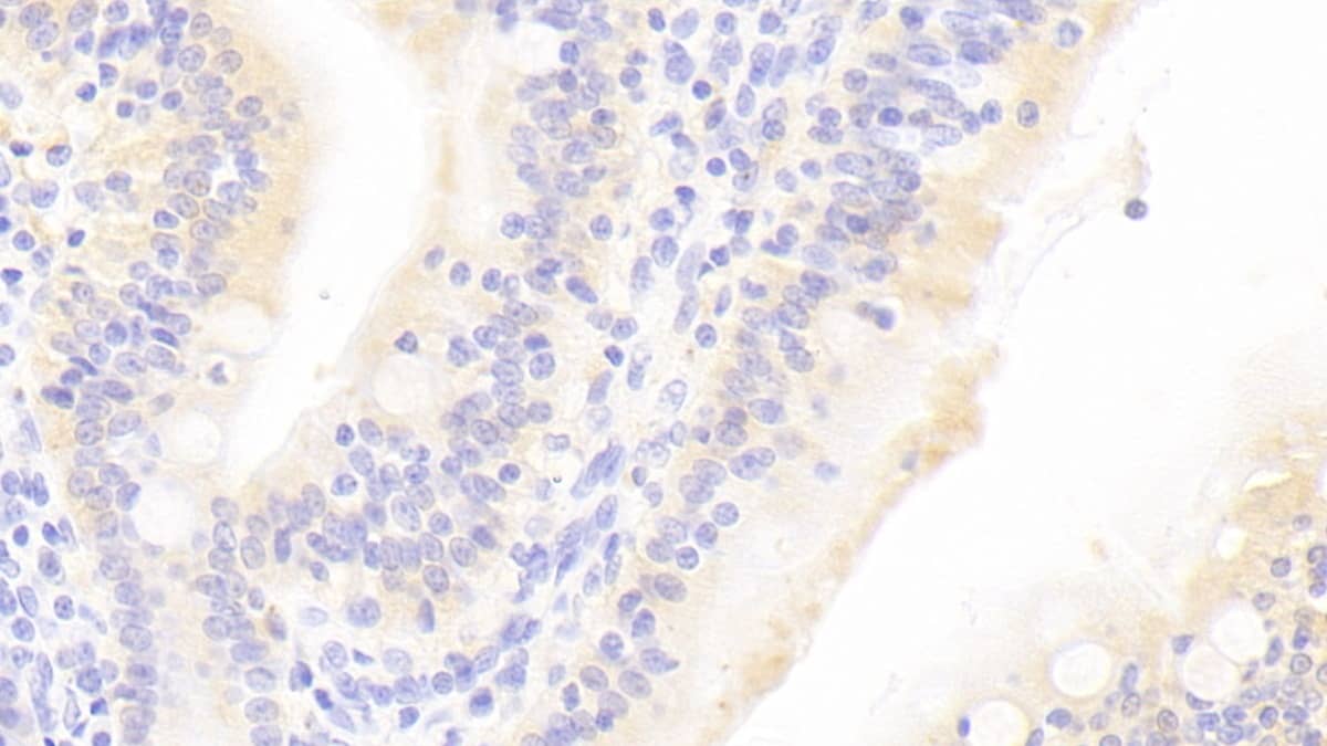
Immunohistochemistry-Paraffin: PAX8 Antibody [NBP1-32440] - Mouse liver. PAX8 antibody diluted at 1:500. Antigen Retrieval: Citrate buffer, pH 6.0, 15 min.
Immunoprecipitation: PAX8 Antibody [NBP1-32440] - Sample: 1000 ug 293T whole cell lysate/extract A. 40 ug 293T whole cell lysate/extract, B. Control with 2 ug of preimmune rabbit IgG, C. Immunoprecipitation of PAX8 ...read more
Immunocytochemistry/ Immunofluorescence: PAX8 Antibody [NBP1-32440] - Differentiation of human iPSC clone IV towards renal commitment.(a) Representative immunofluorescence images of co-staining for Lhx1/Osr1 up to day ...read more
Immunohistochemistry-Paraffin: PAX8 Antibody [NBP1-32440] - PDX model establishment from ccRCC injection. a Hematoxylin & Eosin staining of human tumor engrafted in murine models. A epresentative image of kidney PDX ...read more
Western Blot: PAX8 Antibody [NBP1-32440] - Non-transfected (-) and transfected (+) 293T whole cell extracts (30 ug) were separated by 10% SDS-PAGE, and the membrane was blotted with PAX8 antibody (NBP1-32440) diluted at ...read more
Immunohistochemistry-Paraffin: PAX8 Antibody [NBP1-32440] - PAX8 antibody detects PAX8 protein at nucleus by immunohistochemical analysis.Sample: Paraffin-embedded dog kidney.PAX8 stained by PAX8 antibody (NBP1-32440) ...read more
Western Blot: PAX8 Antibody [NBP1-32440] - Whole cell extract (30 ug) was separated by 10% SDS-PAGE, and the membrane was blotted with PAX8 antibody (NBP1-32440) diluted at 1:500. The HRP-conjugated anti-rabbit IgG ...read more
Product Details
Summary
Reactivity
Hu, Mu, Rt, Ca, XpSpecies Glossary
Applications
WB, Simple Western, ICC/IF, IHC, IP, ChIP, Single-Cell Western
In Simple Western only 10 - 15 uL of the recommended dilution is used per data point. Separated by Size-Wes, Sally Sue/Peggy Sue. SCW is validated using 786-O cells.
Theoretical MW
48 kDa. Disclaimer note: The observed molecular weight of the protein may vary from the listed predicted molecular weight due to post translational modifications, post translation cleavages, relative charges, and other experimental factors.
Reviewed Applications
Read 1 Review rated 4 using NBP1-32440 in the following applications:
Aliquot and store at -20C or -80C. Avoid freeze-thaw cycles.
Buffer
PBS, 20% Glycerol
Preservative
0.025% Proclin 300
Purity
Antigen Affinity-purified
Alternate Names for PAX8 Antibody
paired box 8
paired box gene 8
paired box protein Pax-8
paired domain gene 8
Background
This gene encodes a member of the paired box (PAX) family of transcription factors. Members of this gene family typically encode proteins that contain a paired box domain, an octapeptide, and a paired-type homeodomain. This nuclear protein is involved in thyroid follicular cell development and expression of thyroid-specific genes. Mutations in this gene have been associated with thyroid dysgenesis, thyroid follicular carcinomas and atypical follicular thyroid adenomas. Alternateively spliced transcript variants encoding different isoforms have been described.
Limitations
This product is for research use only and is not approved for use in humans or in clinical diagnosis. Primary Antibodies are guaranteed for 1 year from date of receipt.
Lakshmi Varahan B Investigating the effects of aspirin in high-grade serous ovarian carcinoma models in vitro Thesis 2023-01-01 (Immunohistochemistry-Paraffin, Human)
***Bio-Techne Response: This review was submitted through the legacy Novus Innovators Program, reflecting a new species or application tested on a primary antibody.***
Product General Protocols
Find general support by application which include: protocols, troubleshooting, illustrated assays, videos and webinars.
The concentration calculator allows you to quickly calculate the volume, mass or concentration of your vial. Simply enter your mass, volume, or concentration values for your reagent and the calculator will determine the rest.

![Western Blot: PAX8 Antibody [NBP1-32440] - 293T whole cell and nuclear extracts (30 ug) were separated by 10% SDS-PAGE, and the membrane was blotted with PAX8 antibody diluted at 1:1000.](http://images.novusbio.com/fullsize/PAX8-Antibody-Western-Blot-NBP1-32440-img0019.jpg "Western Blot: PAX8 Antibody [NBP1-32440] - 293T whole cell and nuclear extracts (30 ug) were separated by 10% SDS-PAGE, and the membrane was blotted with PAX8 antibody diluted at 1:1000.")


![Western Blot TSHR Antibody (484405) [Unconjugated]](https://images.novusbio.com/images2/TSH_R_MAB6534_Western_Blot_10858.jpg)
![ELISA TSHR Antibody (484405) [Unconjugated]](https://images.novusbio.com/images2/TSH_R_MAB6534_ELISA_21565.jpg)


![Immunocytochemistry/Immunofluorescence: PAX8 Antibody [NBP1-32440] - Stepwise differentiation of human iPSCs towards renal progenitor cells (RPCs). Schematic description of the two-stage protocol applied to iPSC-renal commitment. Immunofluorescence of iPSCs (derived from retroviral transfected dermal fibroblasts) exposed to differentiating media. Pictured are the IM and MM marker expressions as Wt1, Pax8, Pax2, Six2 and Sall1. Image collected and cropped by CiteAb from the following publication (//www.nature.com/articles/srep08826), licensed under a CC-BY license.](http://images.novusbio.com/fullsize/PAX8-Antibody-Immunocytochemistry-Immunofluorescence-NBP1-32440-img0020.jpg "Immunocytochemistry/Immunofluorescence: PAX8 Antibody [NBP1-32440] - Stepwise differentiation of human iPSCs towards renal progenitor cells (RPCs). Schematic description of the two-stage protocol applied to iPSC-renal commitment. Immunofluorescence of iPSCs (derived from retroviral transfected dermal fibroblasts) exposed to differentiating media. Pictured are the IM and MM marker expressions as Wt1, Pax8, Pax2, Six2 and Sall1. Image collected and cropped by CiteAb from the following publication (//www.nature.com/articles/srep08826), licensed under a CC-BY license.")
![Immunohistochemistry-Paraffin: PAX8 Antibody [NBP1-32440] - Pax8 immunoreactivity in canine renal carcinoma. Primary antibody incubated with tissue for 1h at room temperature. IHC-P image submitted by a verified customer review.](http://images.novusbio.com/fullsize/PAX8-Antibody-Immunohistochemistry-Paraffin-NBP1-32440-img0021.jpg "Immunohistochemistry-Paraffin: PAX8 Antibody [NBP1-32440] - Pax8 immunoreactivity in canine renal carcinoma. Primary antibody incubated with tissue for 1h at room temperature. IHC-P image submitted by a verified customer review.")
![Chromatin Immunoprecipitation: PAX8 Antibody [NBP1-32440] - Sample: 293T whole cell lysate/extract A. 5 ug preimmune rabbit IgG, B. 5 ug of PAX8 antibody The precipitated DNA was detected by PCR with primer set targeting to E2F1 promoter.](http://images.novusbio.com/fullsize/PAX8-Antibody-Chromatin-Immunoprecipitation-NBP1-32440-img0012.jpg "Chromatin Immunoprecipitation: PAX8 Antibody [NBP1-32440] - Sample: 293T whole cell lysate/extract A. 5 ug preimmune rabbit IgG, B. 5 ug of PAX8 antibody The precipitated DNA was detected by PCR with primer set targeting to E2F1 promoter.")
![Immunocytochemistry/Immunofluorescence: PAX8 Antibody [NBP1-32440] - Paraformaldehyde-fixed A549, using antibody at 1:200 dilution.](http://images.novusbio.com/fullsize/PAX8-Antibody-Immunocytochemistry-Immunofluorescence-NBP1-32440-img0011.jpg "Immunocytochemistry/Immunofluorescence: PAX8 Antibody [NBP1-32440] - Paraformaldehyde-fixed A549, using antibody at 1:200 dilution.")
![Immunohistochemistry-Paraffin: PAX8 Antibody [NBP1-32440] - Rat thyroid gland. PAX8 antibody dilution: 1:500. Antigen Retrieval: Trilogy™ (EDTA based, pH 8.0) buffer, 15min.](http://images.novusbio.com/fullsize/PAX8-Antibody-Immunohistochemistry-Paraffin-NBP1-32440-img0015.jpg "Immunohistochemistry-Paraffin: PAX8 Antibody [NBP1-32440] - Rat thyroid gland. PAX8 antibody dilution: 1:500. Antigen Retrieval: Trilogy™ (EDTA based, pH 8.0) buffer, 15min.")
![Immunohistochemistry-Paraffin: PAX8 Antibody [NBP1-32440] - DLD1 xenograft . PAX8 antibody diluted at 1:500. Antigen Retrieval: Trilogy™ (EDTA based, pH 8.0) buffer, 15min.](http://images.novusbio.com/fullsize/PAX8-Antibody-Immunohistochemistry-Paraffin-NBP1-32440-img0016.jpg "Immunohistochemistry-Paraffin: PAX8 Antibody [NBP1-32440] - DLD1 xenograft . PAX8 antibody diluted at 1:500. Antigen Retrieval: Trilogy™ (EDTA based, pH 8.0) buffer, 15min.")
![Immunohistochemistry-Paraffin: PAX8 Antibody [NBP1-32440] - Mouse liver. PAX8 antibody diluted at 1:500. Antigen Retrieval: Citrate buffer, pH 6.0, 15 min.](http://images.novusbio.com/fullsize/PAX8-Antibody-Immunohistochemistry-Paraffin-NBP1-32440-img0018.jpg "Immunohistochemistry-Paraffin: PAX8 Antibody [NBP1-32440] - Mouse liver. PAX8 antibody diluted at 1:500. Antigen Retrieval: Citrate buffer, pH 6.0, 15 min.")
![Immunoprecipitation: PAX8 Antibody [NBP1-32440] - Sample: 1000 ug 293T whole cell lysate/extract A. 40 ug 293T whole cell lysate/extract, B. Control with 2 ug of preimmune rabbit IgG, C. Immunoprecipitation of PAX8 protein by 2 ug of PAX8 antibody 10% SDS-PAGE gel.](http://images.novusbio.com/fullsize/PAX8-Antibody-Immunoprecipitation-NBP1-32440-img0013.jpg "Immunoprecipitation: PAX8 Antibody [NBP1-32440] - Sample: 1000 ug 293T whole cell lysate/extract A. 40 ug 293T whole cell lysate/extract, B. Control with 2 ug of preimmune rabbit IgG, C. Immunoprecipitation of PAX8 protein by 2 ug of PAX8 antibody 10% SDS-PAGE gel.")
![Immunocytochemistry/ Immunofluorescence: PAX8 Antibody [NBP1-32440] - Differentiation of human iPSC clone IV towards renal commitment.(a) Representative immunofluorescence images of co-staining for Lhx1/Osr1 up to day 6. (b) Images of co-staining of Pax2/Six2, Pax8/Six2 & Six2/Wt1 from day 6 to 12. (c) Expression of renal progenitor markers such as CD24, Claudin1 & GGT1 from day 0 to 19. Nuclei are stained with DAPI (blue). Scale bars: 50 μm (a, b), 20 μm (c). (d) Gene expression analysis for renal progenitor markers at different points in time. Image collected & cropped by CiteAb from the following publication (//www.nature.com/articles/srep08826), licensed under a CC-BY license. Not internally tested by Novus Biologicals.](http://images.novusbio.com/fullsize/nbp1-32440_rabbit-polyclonal-pax8-antibody-310202415345341.jpg "Immunocytochemistry/ Immunofluorescence: PAX8 Antibody [NBP1-32440] - Differentiation of human iPSC clone IV towards renal commitment.(a) Representative immunofluorescence images of co-staining for Lhx1/Osr1 up to day 6. (b) Images of co-staining of Pax2/Six2, Pax8/Six2 & Six2/Wt1 from day 6 to 12. (c) Expression of renal progenitor markers such as CD24, Claudin1 & GGT1 from day 0 to 19. Nuclei are stained with DAPI (blue). Scale bars: 50 μm (a, b), 20 μm (c). (d) Gene expression analysis for renal progenitor markers at different points in time. Image collected & cropped by CiteAb from the following publication (//www.nature.com/articles/srep08826), licensed under a CC-BY license. Not internally tested by Novus Biologicals.")
![Immunohistochemistry-Paraffin: PAX8 Antibody [NBP1-32440] - PDX model establishment from ccRCC injection. a Hematoxylin & Eosin staining of human tumor engrafted in murine models. A epresentative image of kidney PDX reported. Microscope Nikon Eclipse E1000 10X & 20X (b) Graph reporting % of patients who engrafted when orthotopically injected in mice & distributed following grading (3 G1; 7 G2; 13 G3; 7 G4). Eight mice for each patient injected & all 18 tumors on 30 which evaluated as engrafted developed tumor masses on ≥ of 6 mice. Tumors declared unable to engraft did not produce, at all, tumor masses. c Representative images (1 × 0.63) of PDXs excised 90 days after injection by Stereomicroscope (Olympus SZX10,XCX50). One representative image for G2, G3 & G4 types reported. d Representative images (1 × 0.63 & 1.25) of aberrant neo-angiogenesis formation in PDXs by Stereomicroscope (Olympus SZX10, XCX50 camera). e Hematoxylin & Eosin staining of PDXs versus parental primary patients. One representative tumor for each grade (G2, G3, G4) reported. Staining executed on OCT frozen samples. f Hematoxylin & Eosin & anti-PAX8, CD10, Vimentin & EMA staining reported on formalin-fixed & paraffin- embedded parental primary, metastatic tissues & PDXs. Primary, metastatic & PDX tissues obtained & shown from one representative patient. Microscope Nikon Eclipse 55i, magnification 20×. g Histogram showing number of engrafting tumor populations evaluated over 30 injected patient samples & correlated w/ patient recurrence frequency calculated as development of metastases after surgery. Orange color represents recurrent (n = 7) & metastatic (n = 3) patients in engrafted group (n = 18). Pink color represents recurrent patients (n = 2) in non-engrafted group (n = 12) for a total of 30 injected samples Image collected & cropped by CiteAb from following publication (//pubmed.ncbi.nlm.nih.gov/30185225), licensed under a CC-BY license. Not internally tested by Novus Biologicals.](http://images.novusbio.com/fullsize/nbp1-32440_rabbit-polyclonal-pax8-antibody-310202416165612.jpg "Immunohistochemistry-Paraffin: PAX8 Antibody [NBP1-32440] - PDX model establishment from ccRCC injection. a Hematoxylin & Eosin staining of human tumor engrafted in murine models. A epresentative image of kidney PDX reported. Microscope Nikon Eclipse E1000 10X & 20X (b) Graph reporting % of patients who engrafted when orthotopically injected in mice & distributed following grading (3 G1; 7 G2; 13 G3; 7 G4). Eight mice for each patient injected & all 18 tumors on 30 which evaluated as engrafted developed tumor masses on ≥ of 6 mice. Tumors declared unable to engraft did not produce, at all, tumor masses. c Representative images (1 × 0.63) of PDXs excised 90 days after injection by Stereomicroscope (Olympus SZX10,XCX50). One representative image for G2, G3 & G4 types reported. d Representative images (1 × 0.63 & 1.25) of aberrant neo-angiogenesis formation in PDXs by Stereomicroscope (Olympus SZX10, XCX50 camera). e Hematoxylin & Eosin staining of PDXs versus parental primary patients. One representative tumor for each grade (G2, G3, G4) reported. Staining executed on OCT frozen samples. f Hematoxylin & Eosin & anti-PAX8, CD10, Vimentin & EMA staining reported on formalin-fixed & paraffin- embedded parental primary, metastatic tissues & PDXs. Primary, metastatic & PDX tissues obtained & shown from one representative patient. Microscope Nikon Eclipse 55i, magnification 20×. g Histogram showing number of engrafting tumor populations evaluated over 30 injected patient samples & correlated w/ patient recurrence frequency calculated as development of metastases after surgery. Orange color represents recurrent (n = 7) & metastatic (n = 3) patients in engrafted group (n = 18). Pink color represents recurrent patients (n = 2) in non-engrafted group (n = 12) for a total of 30 injected samples Image collected & cropped by CiteAb from following publication (//pubmed.ncbi.nlm.nih.gov/30185225), licensed under a CC-BY license. Not internally tested by Novus Biologicals.")
![Western Blot: PAX8 Antibody [NBP1-32440] - Non-transfected (-) and transfected (+) 293T whole cell extracts (30 ug) were separated by 10% SDS-PAGE, and the membrane was blotted with PAX8 antibody (NBP1-32440) diluted at 1:10000. The HRP-conjugated anti-rabbit IgG antibody was used to detect the primary antibody.](http://images.novusbio.com/fullsize/nbp1-32440_rabbit-polyclonal-pax8-antibody-910202420134072.jpg "Western Blot: PAX8 Antibody [NBP1-32440] - Non-transfected (-) and transfected (+) 293T whole cell extracts (30 ug) were separated by 10% SDS-PAGE, and the membrane was blotted with PAX8 antibody (NBP1-32440) diluted at 1:10000. The HRP-conjugated anti-rabbit IgG antibody was used to detect the primary antibody.")
![Immunohistochemistry-Paraffin: PAX8 Antibody [NBP1-32440] - PAX8 antibody detects PAX8 protein at nucleus by immunohistochemical analysis.Sample: Paraffin-embedded dog kidney.PAX8 stained by PAX8 antibody (NBP1-32440) diluted at 1:2000.Antigen Retrieval: Citrate buffer, pH 6.0, 15 min](http://images.novusbio.com/fullsize/nbp1-32440_rabbit-polyclonal-pax8-antibody-910202419593589.jpg "Immunohistochemistry-Paraffin: PAX8 Antibody [NBP1-32440] - PAX8 antibody detects PAX8 protein at nucleus by immunohistochemical analysis.Sample: Paraffin-embedded dog kidney.PAX8 stained by PAX8 antibody (NBP1-32440) diluted at 1:2000.Antigen Retrieval: Citrate buffer, pH 6.0, 15 min")
![Western Blot: PAX8 Antibody [NBP1-32440] - Whole cell extract (30 ug) was separated by 10% SDS-PAGE, and the membrane was blotted with PAX8 antibody (NBP1-32440) diluted at 1:500. The HRP-conjugated anti-rabbit IgG antibody was used to detect the primary antibody.](http://images.novusbio.com/fullsize/nbp1-32440_rabbit-polyclonal-pax8-antibody-9102024201466.jpg "Western Blot: PAX8 Antibody [NBP1-32440] - Whole cell extract (30 ug) was separated by 10% SDS-PAGE, and the membrane was blotted with PAX8 antibody (NBP1-32440) diluted at 1:500. The HRP-conjugated anti-rabbit IgG antibody was used to detect the primary antibody.")


![Immunocytochemistry Pax2 Antibody [Unconjugated]](https://images.novusbio.com/images2/Pax2_AF3364_Immunocytochemistry_14405.jpg)
![Western Blot Pax2 Antibody [Unconjugated]](https://images.novusbio.com/images2/af3364_human-pax2-affinity-purified-polyclonal-ab-western-blot-145202183852.jpg)
![Immunohistochemistry Pax2 Antibody [Unconjugated]](https://images.novusbio.com/images2/af3364_human-pax2-affinity-purified-polyclonal-ab-immunohistochemistry-192021142455.jpg)
![Immunohistochemistry Ret Antibody [Unconjugated]](https://images.novusbio.com/images2/Ret_AF482_Immunohistochemistry_10194.jpg)
![Western Blot Ret Antibody [Unconjugated]](https://images.novusbio.com/images/af482_mouse-ret-affinity-purified-polyclonal-ab-8220249503265.jpg)
![Immunohistochemistry Ret Antibody [Unconjugated]](https://images.novusbio.com/images/af482_mouse-ret-affinity-purified-polyclonal-ab-8220249505961.jpg)


![Immunohistochemistry Patched 1/PTCH Antibody (413220) [Unconjugated] - (First Extracellular Loop)](https://images.novusbio.com/images2/Patched_1_MAB41051_Immunohistochemistry_15456.jpg)
![Western Blot Patched 1/PTCH Antibody (413220) [Unconjugated] - (First Extracellular Loop)](https://images.novusbio.com/images2/Patched_1_MAB41051_Western_Blot_10905.jpg)
![Immunohistochemistry Patched 1/PTCH Antibody (413220) [Unconjugated] - (First Extracellular Loop)](https://images.novusbio.com/images2/Patched_1_MAB41051_Immunohistochemistry_7016.jpg)


![Flow Cytometry Pax5/BSAP Antibody (1207C) [Unconjugated]](https://images.novusbio.com/images2/Pax5_MAB3487_Flow_Cytometry_19328.jpg)
![Western Blot Pax5/BSAP Antibody (1207C) [Unconjugated]](https://images.novusbio.com/images2/Pax5_MAB3487_Western_Blot_19363.jpg)
![Immunohistochemistry Pax5/BSAP Antibody (1207C) [Unconjugated]](https://images.novusbio.com/images2/Pax5_MAB3487_Immunohistochemistry_19379.jpg)
![Bioactivity Thrombopoietin/THPO [Unconjugated]](https://images.novusbio.com/images/protein/288-tpncf_recombinant-human-thrombopoietin-ns0-expressed-protein-cf-bioactivity-272020135047.jpg)
 using a 1:1000 dilution of HRP-conjugated Anti-Rabbit IgG Secondary Antibody (Catalog # HAF008). This experiment was conducted under reducing conditions and using Immunoblot Buffer Group 1.")
![Western Blot: Goat anti-Rabbit IgG (H+L) Secondary Antibody [HRP] [NB7160] - Western blot showing vemurafenib treatment in BRAFV600E CRC cells inhibits fission mediator DRP1 with no significant effect on fusion proteins (Mfn1 & 2) using MFN-1 antibody (NBP1-51841) and corresponding secondary antibody, goat anti-rabbit IgG-HRP (NB7160). Image collected and cropped by CiteAb from the following publication (https://pubmed.ncbi.nlm.nih.gov/33738242).](https://images.novusbio.com/images/Goat-anti-Rabbit-IgG-H+L-Secondary-Antibody-HRP-Western-Blot-NB7160-img0001.jpg "Western Blot: Goat anti-Rabbit IgG (H+L) Secondary Antibody [HRP] [NB7160] - Western blot showing vemurafenib treatment in BRAFV600E CRC cells inhibits fission mediator DRP1 with no significant effect on fusion proteins (Mfn1 & 2) using MFN-1 antibody (NBP1-51841) and corresponding secondary antibody, goat anti-rabbit IgG-HRP (NB7160). Image collected and cropped by CiteAb from the following publication (https://pubmed.ncbi.nlm.nih.gov/33738242).")
![Flow Cytometry: Rabbit IgG Isotype Control [NBP2-24891] - Intracellular FACS analysis of mouse TLR6 polyclonal antibody (red), rabbit isotype control (green), RAW cells alone (shaded). Two micrograms of antibodies were used. Goat anti-rabbit FITC (Novus, 20302) was used as secondary.](https://images.novusbio.com/images/Rabbit--Mouse-IgG-Isotype-Control-Flow-Cytometry-NBP2-24891-img0001.jpg "Flow Cytometry: Rabbit IgG Isotype Control [NBP2-24891] - Intracellular FACS analysis of mouse TLR6 polyclonal antibody (red), rabbit isotype control (green), RAW cells alone (shaded). Two micrograms of antibodies were used. Goat anti-rabbit FITC (Novus, 20302) was used as secondary.")
