
![Western Blot: Cytokeratin, pan Antibody (PCK-26) [NB120-6401] - Whole extract of HeLa cells was separated on SDS-PAGE and probed with Monoclonal Anti-Cytokeratin, pan Clone: PCK-26. WB was developed using Goat Anti-Mouse IgG-Peroxidase and a chemiluminescent substrate. Lane 1: Antibody dilution 1:300. Lane 2: Antibody dilution 1:500. Lane 3: Antibody dilution 1:1000.](http://images.novusbio.com/fullsize/pan-Cytokeratin-Antibody-PCK-26-Western-Blot-NB120-6401-img0011.jpg "Western Blot: Cytokeratin, pan Antibody (PCK-26) [NB120-6401] - Whole extract of HeLa cells was separated on SDS-PAGE and probed with Monoclonal Anti-Cytokeratin, pan Clone: PCK-26. WB was developed using Goat Anti-Mouse IgG-Peroxidase and a chemiluminescent substrate. Lane 1: Antibody dilution 1:300. Lane 2: Antibody dilution 1:500. Lane 3: Antibody dilution 1:1000.")
| Reactivity | Hu, Mu, Rt, Po, Bv, Ca, Ch, Eq, Fe, Fi, Gp, Gt, Ha, Rb, Re, ShSpecies Glossary |
| Applications | WB, ICC/IF, IHC |
| Clone | PCK-26 |
| Clonality | Monoclonal |
| Host | Mouse |
| Conjugate | Unconjugated |
| Immunogen | This Cytokeratin, pan Antibody (PCK-26) was developed against the full length native protein (purified) (Human epidermis). |
| Localization | Cytoplasmic |
| Marker | Epithelial marker |
| Specificity | PCK-26 recognizes the 58 kDa cytokeratin 5, the 56 kDa cytokeratin 6 and the 52 kDa cytokeratin 8 band in immunoblotting (Lane EB et al). The antibody recognizes an epitope located on the Type II cytokeratins 1, 5, 6, and 8. PCK-26 is a broad spectrum antibody which reacts specifically with a variety of normal, reactive, and neoplastic epithelial tissues. The antibody reacts with simple, cornifying, and non-cornifying squamous epithelia and pseudostratified epithelia. |
| Isotype | IgG1 |
| Clonality | Monoclonal |
| Host | Mouse |
| Gene | KRT1 |
| Purity | Unpurified |
| Innovator's Reward | Test in a species/application not listed above to receive a full credit towards a future purchase. |
| Dilutions |
|
|
| Reviewed Applications |
|
|
| Publications |
|
| Storage | Store at 4C short term. Aliquot and store at -20C long term. Avoid freeze-thaw cycles. |
| Buffer | Ascites |
| Preservative | 0.09% Sodium Azide |
| Purity | Unpurified |


![Choline Kinase beta [Unconjugated]](/sites/all/modules/enterprise-tech/et_datasheets/images/novus_guarantee.png "Choline Kinase beta [Unconjugated]")


![Simple Western Albumin Antibody (188835) [Unconjugated] - Serum](https://images.novusbio.com/images2/Albumin_MAB1455_Simple_Western_21888.jpg)
![Immunocytochemistry Albumin Antibody (188835) [Unconjugated] - Serum](https://images.novusbio.com/images2/Albumin_MAB1455_Immunocytochemistry__Immunofluorescence_17777.jpg)
![Immunohistochemistry Albumin Antibody (188835) [Unconjugated] - Serum](https://images.novusbio.com/images2/Albumin_MAB1455_Immunohistochemistry_23317.jpg)


| Images | Ratings | Applications | Species | Date | Details | ||||||||
|---|---|---|---|---|---|---|---|---|---|---|---|---|---|
Enlarge |
reviewed by:
Verified Customer |
ICC | Human | 08/09/2019 |
Summary
|
||||||||
Enlarge |
reviewed by:
Abhishek Aggarwal |
IF | Human | 02/05/2013 |
Summary
|
Secondary Antibodies |
Isotype Controls |
Research Areas for Cytokeratin, pan Antibody (NB120-6401)Find related products by research area.
|

The concentration calculator allows you to quickly calculate the volume, mass or concentration of your vial. Simply enter your mass, volume, or concentration values for your reagent and the calculator will determine the rest.
5 | |
4 | |
3 | |
2 | |
1 |
| Verified Customer 08/09/2019 |
||
| Application: | ICC | |
| Species: | Human |
| Abhishek Aggarwal 02/05/2013 |
||
| Application: | IF | |
| Species: | Human |


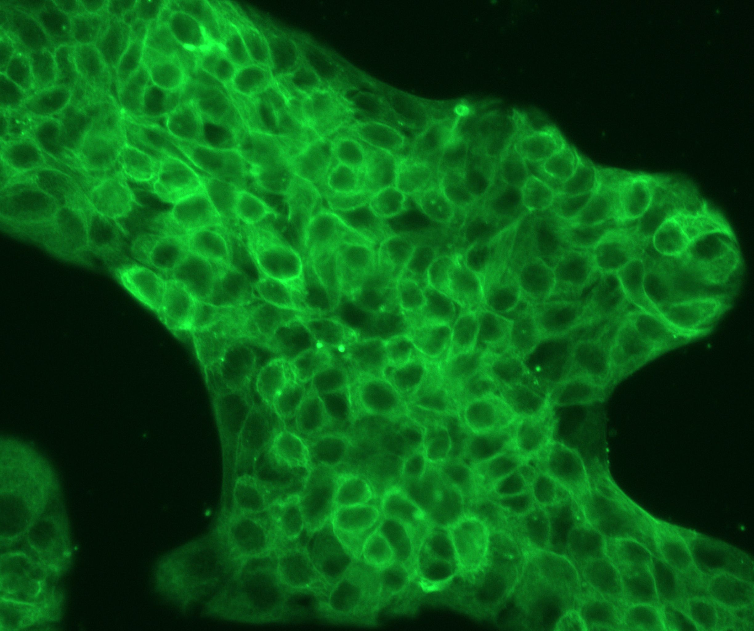
![Immunocytochemistry/Immunofluorescence: Cytokeratin, pan Antibody (PCK-26) [NB120-6401] - Human pancreas cancer cells. Antibody at 1:250 dilution. Overnight incubation. No permeabilization. ICC/IF image submitted by a verified customer review.](http://images.novusbio.com/fullsize/Cytokeratin-pan-Antibody-PCK-26-Immunocytochemistry-Immunofluorescence-NB120-6401-img0012.jpg "Immunocytochemistry/Immunofluorescence: Cytokeratin, pan Antibody (PCK-26) [NB120-6401] - Human pancreas cancer cells. Antibody at 1:250 dilution. Overnight incubation. No permeabilization. ICC/IF image submitted by a verified customer review.")
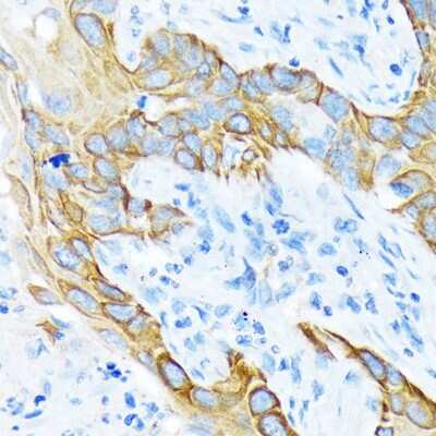
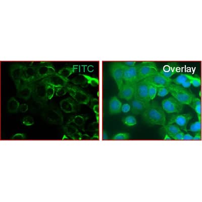
![Immunohistochemistry-Paraffin: Cytokeratin, pan Antibody (PCK-26) [NB120-6401] - Staining of FFPE human placenta sections with 1:300 Monoclonal Anti-Cytokeratin, pan Clone: PCK-26, followed by Goat Anti Mouse IgG (Fab)-FITC.](http://images.novusbio.com/fullsize/pan-Cytokeratin-Antibody-PCK-26-Immunohistochemistry-Paraffin-NB120-6401-img0006.jpg "Immunohistochemistry-Paraffin: Cytokeratin, pan Antibody (PCK-26) [NB120-6401] - Staining of FFPE human placenta sections with 1:300 Monoclonal Anti-Cytokeratin, pan Clone: PCK-26, followed by Goat Anti Mouse IgG (Fab)-FITC.")
![Immunohistochemistry: Cytokeratin, pan Antibody (PCK-26) [NB120-6401] - Histological examination of the subcutaneous xenografts in nude mice with H&E staining (left panel) and IHC with anti-pan CK (right panel) at 400x magnification. Asterisks (*) indicate representative keratinization and keratin-pearl formation. Image collected and cropped by CiteAb from the following publication (mdpi.com/2072-6694/12/1/61), licensed under a CC-BY license.](http://images.novusbio.com/fullsize/Cytokeratin-pan-Antibody-PCK-26-Immunohistochemistry-NB120-6401-img0014.jpg "Immunohistochemistry: Cytokeratin, pan Antibody (PCK-26) [NB120-6401] - Histological examination of the subcutaneous xenografts in nude mice with H&E staining (left panel) and IHC with anti-pan CK (right panel) at 400x magnification. Asterisks (*) indicate representative keratinization and keratin-pearl formation. Image collected and cropped by CiteAb from the following publication (mdpi.com/2072-6694/12/1/61), licensed under a CC-BY license.")
![Immunohistochemistry: Cytokeratin, pan Antibody (PCK-26) [NB120-6401] - Histological examination of NHRI-HN1 tumors in nude mice and B6 mice with H&E staining (upper panels) at 400x and 1000x magnifications and IHC with anti-pan CK (middle panel) and EGFR (lower panel) at 400x magnification. Image collected and cropped by CiteAb from the following publication (mdpi.com/2072-6694/12/1/61), licensed under a CC-BY license.](http://images.novusbio.com/fullsize/Cytokeratin-pan-Antibody-PCK-26-Immunohistochemistry-NB120-6401-img0013.jpg "Immunohistochemistry: Cytokeratin, pan Antibody (PCK-26) [NB120-6401] - Histological examination of NHRI-HN1 tumors in nude mice and B6 mice with H&E staining (upper panels) at 400x and 1000x magnifications and IHC with anti-pan CK (middle panel) and EGFR (lower panel) at 400x magnification. Image collected and cropped by CiteAb from the following publication (mdpi.com/2072-6694/12/1/61), licensed under a CC-BY license.")
![Immunocytochemistry/ Immunofluorescence: Cytokeratin, pan Antibody (PCK-26) [NB120-6401] - Detection of EMT & increased motility in NHRI-HN1 cells. (A) Summary of the most enriched pathways associated with tumorigenesis in syngeneic mice by comparing tumorigenic NHRI-HN1 cells with nontumorigenic cells, including M1-2, M2-3 & NHRI-HN2, using GSEA analysis. Blue indicates a negative normalized enrichment score (NES) & orange indicates a positive NES. (B) Morphology of M1-2 & NHRI-HN1 cells via phase-contrast microscopy at 100× magnification. (C) Relative adhesion activity in M1-2 & NHRI-HN1 cells, determined by normalizing the mean OD 490 nm value of NHRI-HN1 cells to that of M1-2 cells. (D) Cells stained with Alexa Fluor 488 phalloidin, anti-pan-CK & anti-EGFR antibodies at 400× magnification. (E) Immunoblot analysis of epithelial (E-cadherin, alpha -catenin & beta -catenin), mesenchymal (N-cadherin & Vimentin) proteins & EMT-related transcription factors, including Twist, Snail & Slug in M1-2 & NHRI-HN1 cells. Protein levels were normalized to an internal control, alpha -tubulin. Ratios were determined by dividing the normalized protein levels in NHRI-HN1 cells by that in M1-2 cells. (F) Representative images (left) & relative data (right) for migration activity of M1-2 & NHRI-HN1 cells. (G) Representative images (left) & relative data (right) for invasion activity of M1-2 & NHRI-HN1 cells at 200× magnification. The relative migration or invasion activity was determined by normalizing the mean number of cells that have migrated or invaded per field of NHRI-HN1 cells to that of M1-2 cells. Error bars represent SE; ** p < 0.01; *** p < 0.001. Image collected & cropped by CiteAb from the following publication (//pubmed.ncbi.nlm.nih.gov/31878324), licensed under a CC-BY license. Not internally tested by Novus Biologicals.](http://images.novusbio.com/fullsize/nb120-6401_mouse-monoclonal-cytokeratin-pan-antibody-pck-26-310202416212383.jpg "Immunocytochemistry/ Immunofluorescence: Cytokeratin, pan Antibody (PCK-26) [NB120-6401] - Detection of EMT & increased motility in NHRI-HN1 cells. (A) Summary of the most enriched pathways associated with tumorigenesis in syngeneic mice by comparing tumorigenic NHRI-HN1 cells with nontumorigenic cells, including M1-2, M2-3 & NHRI-HN2, using GSEA analysis. Blue indicates a negative normalized enrichment score (NES) & orange indicates a positive NES. (B) Morphology of M1-2 & NHRI-HN1 cells via phase-contrast microscopy at 100× magnification. (C) Relative adhesion activity in M1-2 & NHRI-HN1 cells, determined by normalizing the mean OD 490 nm value of NHRI-HN1 cells to that of M1-2 cells. (D) Cells stained with Alexa Fluor 488 phalloidin, anti-pan-CK & anti-EGFR antibodies at 400× magnification. (E) Immunoblot analysis of epithelial (E-cadherin, alpha -catenin & beta -catenin), mesenchymal (N-cadherin & Vimentin) proteins & EMT-related transcription factors, including Twist, Snail & Slug in M1-2 & NHRI-HN1 cells. Protein levels were normalized to an internal control, alpha -tubulin. Ratios were determined by dividing the normalized protein levels in NHRI-HN1 cells by that in M1-2 cells. (F) Representative images (left) & relative data (right) for migration activity of M1-2 & NHRI-HN1 cells. (G) Representative images (left) & relative data (right) for invasion activity of M1-2 & NHRI-HN1 cells at 200× magnification. The relative migration or invasion activity was determined by normalizing the mean number of cells that have migrated or invaded per field of NHRI-HN1 cells to that of M1-2 cells. Error bars represent SE; ** p < 0.01; *** p < 0.001. Image collected & cropped by CiteAb from the following publication (//pubmed.ncbi.nlm.nih.gov/31878324), licensed under a CC-BY license. Not internally tested by Novus Biologicals.")


![N/A Endostatin [HRP]](https://images.novusbio.com/images2/DATA_Endostatin_DNST0_ELISA_778.jpg)
![N/A Endostatin [HRP]](https://images.novusbio.com/images2/Endostatin_DNST0_ELISA_152.jpg)


 using a 1:1000 dilution of HRP-conjugated Anti-Mouse IgG Secondary Antibody (Catalog # HAF007). This experiment was conducted under reducing conditions and using Immunoblot Buffer Group 1.")
![SDS-Page: Mouse IgG1 Isotype Control (MG1) [NBP1-97005] - Lane 1: , Non-reduced. M: Opal Pre-stained Ladder. Lane 2: , Reduced. Load: 1.0 ug per lane. Predicted/Observed: 120 kDa Non-reduced, 55 and 25 Reduced.](https://images.novusbio.com/images/Mouse-IgG1-Isotype-Control-MG1-SDS-Page-NBP1-97005-img0002.jpg "SDS-Page: Mouse IgG1 Isotype Control (MG1) [NBP1-97005] - Lane 1: , Non-reduced. M: Opal Pre-stained Ladder. Lane 2: , Reduced. Load: 1.0 ug per lane. Predicted/Observed: 120 kDa Non-reduced, 55 and 25 Reduced.")

