| Submit your blog on Tear Secretion to be featured! |
| Submit your event on Tear Secretion to be featured! |
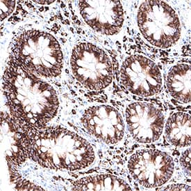
![Immunohistochemistry-Paraffin: LACRT Antibody [NBP3-41240] - Used in DAB staining on fromalin fixed paraffin-embedded Liver tissue](https://images.novusbio.com/fullsize/nbp3-41240_rabbit-lacrt-pab-612202421373087.jpg)
![Western Blot: LACRT Antibody [NBP3-41240] - Sample: Recombinant LACRT, Human.](https://images.novusbio.com/fullsize/nbp3-41240_rabbit-lacrt-pab-612202421195661.jpg)
Rabbit Polyclonal
Species Human, Mouse
Applications WB
|
|
Mouse Monoclonal
Species Human
Applications WB, Simple Western, IHC
|
|
![Immunohistochemistry-Paraffin: Aquaporin-4 Antibody [NBP1-87679] - Analysis in human cerebral cortex and liver tissues. Corresponding AQP4 RNA-seq data are presented for the same tissues.](https://images.novusbio.com/fullsize/Aquaporin-4-Antibody-Immunohistochemistry-Paraffin-NBP1-87679-img0027.jpg)
![Western Blot: Aquaporin-4 Antibody [NBP1-87679] - Analysis in human brain tissue.](https://images.novusbio.com/fullsize/Aquaporin-4-Antibody-Western-Blot-NBP1-87679-img0017.jpg)
Rabbit Polyclonal
Species Human, Mouse, Rat
Applications WB, IHC, IHC-Fr
| 2 Reviews 21 Publications |
|


Rat Monoclonal
Species Human, Mouse, Bovine
Applications IHC, CyTOF-ready, ICC
| 25 Publications |
|

Goat Polyclonal
Species Human
Applications WB
|
|

Mouse Monoclonal
Species Human
Applications WB, IHC
| 2 Publications |
|


Mouse Monoclonal
Species Human
Applications WB, Simple Western, IHC
| 3 Reviews 54 Publications |
|
![Immunohistochemistry-Paraffin: Aquaporin-5 Antibody [NBP2-39043] - Analysis in human salivary gland and liver tissues. Corresponding AQP5 RNA-seq data are presented for the same tissues.](https://images.novusbio.com/fullsize/Aquaporin-5-Antibody-Immunohistochemistry-Paraffin-NBP2-39043-img0006.jpg)
![Immunocytochemistry/Immunofluorescence: Aquaporin-5 Antibody [NBP2-39043] - Staining of human cell line SiHa shows localization to plasma membrane. Antibody staining is shown in green.](https://images.novusbio.com/fullsize/Aquaporin-5-Antibody-Immunocytochemistry-Immunofluorescence-NBP2-39043-img0002.jpg)
Rabbit Polyclonal
Species Human
Applications ICC/IF, IHC, IHC-P
| 1 Publication |
|
![N/A Serpin F1/PEDF [Biotin]](https://images.novusbio.com/fullsize2/DATA_Serpin_F1_DY117705_ELISA_2344.jpg)
Species Human
Applications ELISA
| 19 Publications |
|
![N/A Substance P [Biotin]](https://images.novusbio.com/fullsize2/DATA_Substance_P_KGE007_ELISA_909.jpg)
![N/A Substance P [Biotin]](https://images.novusbio.com/fullsize2/Substance_P_KGE007_ELISA_408.jpg)
Species Multi-Species
Applications ELISA
| 38 Publications |
|


Species Human
Applications BA
| 3 Reviews 820 Publications |
|


Species Human
Applications BA
| 60 Publications |
|
![N/A AGER [HRP]](https://images.novusbio.com/fullsize2/DATA_RAGE_DRG00_ELISA_806.jpg)
![N/A AGER [HRP]](https://images.novusbio.com/fullsize2/DATA_RAGE_DRG00_ELISA_807.jpg)
Species Human
Applications ELISA
| 82 Publications |
|
![Western Blot: CLN3 Antibody [NBP2-43782] - Analysis of A. 30 ug NT2D1 whole cell lysate/extract 10 % SDS-PAGE CLN3 antibody dilution: 1:1000](https://images.novusbio.com/fullsize/CLN3-Antibody-Western-Blot-NBP2-43782-img0002.jpg)
![Western Blot: CLN3 Antibody [NBP2-43782] - Analysis of A. 30 ug Jurkat whole cell lysate/extract B. 30 ug Raji whole cell lysate/extract C. 30 ug NCI-H929 whole cell lysate/extract 10 % SDS-PAGE CLN3 antibody dilution: 1:1000.](https://images.novusbio.com/fullsize/CLN3-Antibody-Western-Blot-NBP2-43782-img0001.jpg)
Rabbit Polyclonal
Species Human
Applications WB
| 1 Publication |
|
![Western Blot: VIP Antibody (1E8) [NBP2-46349] - Analysis of HEK293T cells were transfected with the pCMV6-ENTRY control (Left lane) or pCMV6-ENTRY VIP.](https://images.novusbio.com/fullsize/VIP-Antibody-1E8-Western-Blot-NBP2-46349-img0008.jpg)
![Immunohistochemistry: VIP Antibody (1E8) [NBP2-46349] - Analysis of Carcinoma of Human prostate tissue. (Heat-induced epitope retrieval by 1 mM EDTA in 10mM Tris, pH8.5, 120C for 3min)](https://images.novusbio.com/fullsize/VIP-Antibody-1E8-Immunohistochemistry-NBP2-46349-img0007.jpg)
Mouse Monoclonal
Species Human
Applications WB, IHC, IHC-P
|
|
![Immunocytochemistry/Immunofluorescence: Lysozyme Antibody [NBP2-61118] - NIH3T3 cells were fixed and permeabilized for 10 minutes using -20C MeOH. The cells were incubated with anti-Lysozyme NBP2-61118 at 2 ug/ml overnight at 4C and detected with an anti-rabbit Dylight 488 (Green) at a 1:1000 dilution for 60 minutes. Nuclei were counterstained with DAPI (Blue). Cells were imaged using a 100X objective and digitally deconvolved.](https://images.novusbio.com/fullsize/Lysozyme-Antibody-Immunocytochemistry-Immunofluorescence-NBP2-61118-img0006.jpg)
![Western Blot: Lysozyme Antibody [NBP2-61118] - Total protein from human cell lines THP-1 and HepG2, human stomach and kidney as well as mouse kidney was separated on a 4-20% gel by SDS-PAGE, transferred to 0.2 um PVDF membrane and blocked in 5% non-fat milk in TBST. The membrane was probed with 2.0 ug/ml anti-Lysozyme in 5% non-fat milk in TBST and detected with an anti-rabbit HRP secondary antibody using chemiluminescence.](https://images.novusbio.com/fullsize/Lysozyme-Antibody-Western-Blot-NBP2-61118-img0003.jpg)
Rabbit Polyclonal
Species Human, Mouse
Applications WB, Simple Western, Flow
| 7 Publications |
|
![Immunohistochemistry-Paraffin: Adenine Nucleotide Translocator 2 Antibody [NBP2-92630] - Paraffin-embedded rat heart using Adenine Nucleotide Translocator 2 .](https://images.novusbio.com/fullsize/Adenine-Nucleotide-Translocator-2-Antibody-Immunohistochemistry-Paraffin-NBP2-92630-img0003.jpg)
![Immunohistochemistry-Paraffin: Adenine Nucleotide Translocator 2 Antibody [NBP2-92630] - Paraffin-embedded mouse heart using Adenine Nucleotide Translocator 2 .](https://images.novusbio.com/fullsize/Adenine-Nucleotide-Translocator-2-Antibody-Immunohistochemistry-Paraffin-NBP2-92630-img0002.jpg)
Rabbit Polyclonal
Species Human, Mouse, Rat
Applications WB, ELISA, ICC/IF
|
|
