| Submit your blog on Mesodermal Cell Migration to be featured! |
| Submit your event on Mesodermal Cell Migration to be featured! |
![Western Blot: NOD2 Antibody (2D9) [NB100-524] - HCMV infection induces NOD2 mRNA and protein in HFFs and U373 cells. E. U373 glioma cells were infected with HCMV Towne strain and levels of NOD1, NOD2 and GAPDH mRNAs were measured by qRT-PCR at indicated time points. F. HFFs were infected with HCMV (Towne) at MOI of 1 PFU/cell and levels of NOD2 protein and B-actin were determined 48 and 72 hpi. G. HFFs were infected with HCMV (Towne) strain at MOI of 0.03 or 3 PFU/cell and levels of NOD2 protein and B-actin were determined at 48 hpi. Quantitative data represent mean values (+/-SD) of triplicate determinations from three independent experiments (*p<0.05, **p<0.01, ***p<0.001, one-way ANOVA test). Image collected and cropped by CiteAb from the following publication (//doi.org/10.1371/journal.pone.0092704.g001) licensed under a CC-BY license.](https://images.novusbio.com/fullsize/NOD2-Antibody-2D9-Western-Blot-NB100-524-img0018.jpg)
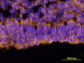
![Immunohistochemistry-Frozen: NOD2 Antibody (2D9) [NB100-524] - Overlay of NOD2-DyLight 488 (green) with phase contrast of murine colon. Image from verified customer review.](https://images.novusbio.com/fullsize/NOD2-Antibody-2D9-Immunohistochemistry-Frozen-NB100-524-img0016.jpg)
Mouse Monoclonal
Species Human, Mouse
Applications WB, Flow, ICC/IF
| 1 Review 26 Publications |
|
![Western Blot: CHIC2 Antibody [NBP1-32719] - Sample (30 ug of whole cell lysate)A: Raji 12% SDS PAGE, antibody diluted at 1:1000.](https://images.novusbio.com/fullsize/CHIC2-Antibody-Western-Blot-NBP1-32719-img0003.jpg)
![Immunocytochemistry/Immunofluorescence: CHIC2 Antibody [NBP1-32719] - Analysis of methanol-fixed HeLa, using CHIC2 antibody (Green) at 1:500 dilution. Alpha-tubulin filaments were labeled with an alpha Tubulin antibody (Red) at 1:2000.](https://images.novusbio.com/fullsize/CHIC2-Antibody-Immunocytochemistry-Immunofluorescence-NBP1-32719-img0002.jpg)
Rabbit Polyclonal
Species Human
Applications WB, ICC/IF
|
|
![Western Blot: Fibronectin Antibody - BSA Free [NBP1-91258] - VSOP observed in perivascular-restricted spinal cord lesions with intact BBB. Immunostaining for laminin (brown) shows vascular endothelium and glia limitans of a perivascular lesion, along with infiltrating cells and VSOP (blue). Image collected and cropped by CiteAb from the following publication (https://asn.sagepub.com/lookup/doi/10.1042/AN20120081), licensed under a CC-BY license.](https://images.novusbio.com/fullsize/Fibronectin-Antibody---BSA-Free-Western-Blot-NBP1-91258-img0020.jpg)
![Immunocytochemistry/Immunofluorescence: Fibronectin Antibody - BSA Free [NBP1-91258] - NIH3T3 cells were fixed in 4% paraformaldehyde for 10 minutes and permeabilized in 0.05% Triton X-100 in PBS for 5 minutes. The cells were incubated with anti- NBP1-91258 at 1 ug/ml overnight at 4C and detected with an anti-rabbit Dylight 488 (Green) at a 1:1000 dilution for 60 minutes. Nuclei were counterstained with DAPI (Blue). Cells were imaged using a 100X objective and digitally deconvolved.](https://images.novusbio.com/fullsize/Fibronectin-Antibody---BSA-Free-Immunocytochemistry-Immunofluorescence-NBP1-91258-img0021.jpg)
Rabbit Polyclonal
Species Human, Mouse, Rat
Applications WB, Simple Western, ICC/IF
| 11 Reviews 56 Publications |
|
![Immunohistochemistry: Rhodopsin Antibody (B630) [NBP2-25160] - Rhodopsin expression and the prevalence of rod photoreceptors in the canine retina. (A) Fluorescence micrographs showing rhodopsin expression (red) in the ABCA4+/+ (left), ABCA4+/- (middle), and ABCA4-/- (right) rod outer segments. Scale bar = 10 um. Image collected and cropped by CiteAb from the following publication (doi.org/10.1371/journal.pgen.1007873) licensed under a CC-BY license.](https://images.novusbio.com/fullsize/Rhodopsin-Antibody-B630-Immunohistochemistry-NBP2-25160-img0004.jpg)
![Western Blot: Rhodopsin Antibody (B630) [NBP2-25160] - Western blot analyses of ABCA4 (above), GAPDH (middle), and RHO (below) protein levels in retinal tissue of dogs with the three different genotypes. Image collected and cropped by CiteAb from the following publication. 50 ug of protein samples were resolved by SDS-PAGE, transferred to nitrocellulose membrane, and immunoblotted with the following primary antibodies: ABCA4 (NBP1-30032, 1:1000) and Rhodopsin (NBP2-25160, 1:5000), followed by Anti-Mouse IgG horseradish peroxidase-conjugated secondary antibody (R&D Systems, HAF007, 1:5000. Image collected and cropped by CiteAb from the following publication (doi.org/10.1371/journal.pgen.1007873) licensed under a CC-BY license.](https://images.novusbio.com/fullsize/Rhodopsin-Antibody-B630-Western-Blot-NBP2-25160-img0005.jpg)
Mouse Monoclonal
Species Human, Mouse, Rat
Applications WB, ICC/IF, IHC
| 10 Publications |
|

Sheep Polyclonal
Species Mouse
Applications WB, IHC
|
|

Sheep Polyclonal
Species Human
Applications WB
| 1 Publication |
|


Mouse Monoclonal
Species Human
Applications WB, Simple Western, IHC
| 2 Publications |
|


Goat Polyclonal
Species Human
Applications WB, Simple Western, Flow
| 30 Publications |
|


Rat Monoclonal
Species Human
Applications WB, Flow, IHC
| 2 Publications |
|


Mouse Monoclonal
Species Human
Applications WB, Flow, IHC
| 1 Review 3 Publications |
|
![Immunocytochemistry/Immunofluorescence: ZFPM1 Antibody [NBP2-47572] - Immunofluorescent staining of human cell line SH-SY5Y shows localization to nucleoplasm, cytosol & vesicles.](https://images.novusbio.com/fullsize/ZFPM1-Antibody-Immunocytochemistry-Immunofluorescence-NBP2-47572-img0003.jpg)
![Immunohistochemistry-Paraffin: ZFPM1 Antibody [NBP2-47572] - Staining of human stomach shows high expression.](https://images.novusbio.com/fullsize/ZFPM1-Antibody-Immunohistochemistry-Paraffin-NBP2-47572-img0005.jpg)
Rabbit Polyclonal
Species Human
Applications ICC/IF, IHC, IHC-P
|
|
![Western Blot: Smad5 [p Ser463, p Ser465] Antibody (SY09-03) [NBP2-67394] - Analysis of Phospho-Smad5(S463/465) on different lysates using anti-Phospho-Smad5(S463/465) antibody at 1/1,000 dilution. Positive control: Lane 1: Mouse brain Lane 2: Rat brain](https://images.novusbio.com/fullsize/Smad5-p-Ser463-p-Ser465-Antibody-SY09-03-Western-Blot-NBP2-67394-img0008.jpg)
![Immunocytochemistry/Immunofluorescence: Smad5 [p Ser463, p Ser465] Antibody (SY09-03) [NBP2-67394] - Staining Phospho-Smad5(S463/465) in SKOV-3 cells (green). The nuclear counter stain is DAPI (blue). Cells were fixed in paraformaldehyde, permeabilised with 0.25% Triton X100/PBS.](https://images.novusbio.com/fullsize/Smad5-p-Ser463-p-Ser465-Antibody-SY09-03-Immunocytochemistry-Immunofluorescence-NBP2-67394-img0002.jpg)
Rabbit Monoclonal
Species Human, Mouse, Rat
Applications WB, ICC/IF, IHC
| 1 Publication |
|
![Immunohistochemistry-Paraffin: PTGER4/EP4 Antibody [NLS3890] - Colon, Carcinoma](https://images.novusbio.com/fullsize/PTGER4-EP4-Antibody-Immunohistochemistry-Paraffin-NLS3890-img0010.jpg)
![Immunohistochemistry-Paraffin: PTGER4/EP4 Antibody [NLS3890] - Analysis of anti-PTGER4 / EP4 antibody with human colon, ganglion at 2 ug/ml.](https://images.novusbio.com/fullsize/PTGER4-EP4-Antibody-Immunohistochemistry-Paraffin-NLS3890-img0003.jpg)
Rabbit Polyclonal
Species Human, Bovine, Monkey
Applications IHC, IHC-P
| 2 Publications |
|


Mouse Monoclonal
Species Human
Applications WB, Flow, CyTOF-ready
|
|
![Western Blot: FGF R1 Antibody (M19B2) [NB600-1287] - Analysis of MCF7 (A), HepG2 (B), NIH3T3 (C), and PC-12 (D) cell lysate using FGF R1 antibody at 2 ug/ml.](https://images.novusbio.com/fullsize/FGF-R1-Antibody-M19B2-Western-Blot-NB600-1287-img0007.jpg)
![Flow Cytometry: FGF R1 Antibody (M19B2) [NB600-1287] - Analysis using the Alexa Fluor 488 (R) conjugate of NB600-1287. Staining of FGF R1 in HEK293 and HEK293 FGFR transfected cells using anti-FGF R1 antibody. (Red) Positive cells, (Green) Negative cells, (Blue) Negative cells + antibody, (Pink) Positive cells + antibody. Image from verified customer review.](https://images.novusbio.com/fullsize/FGF-R1-Antibody-M19B2-Flow-Cytometry-NB600-1287-img0009.jpg)
Mouse Monoclonal
Species Human, Mouse, Rat
Applications WB, Flow, ICC/IF
| 1 Review 7 Publications |
|
![Western Blot: Muscleblind-like 1 Antibody (3E7) [H00004154-M02] - MBNL1 monoclonal antibody (M02), clone 3E7 Analysis of MBNL1 expression in Hela S3 NE.](https://images.novusbio.com/fullsize/Muscleblind-like-1-Antibody-3E7-Western-Blot-H00004154-M02-img0010.jpg)
![Immunocytochemistry/Immunofluorescence: Muscleblind-like 1 Antibody (3E7) [H00004154-M02] - Analysis of monoclonal antibody to MBNL1 on HeLa cell. Antibody concentration 10 ug/ml.](https://images.novusbio.com/fullsize/Muscleblind-like-1-Antibody-3E7-Immunocytochemistry-Immunofluorescence-H00004154-M02-img0006.jpg)
Mouse Monoclonal
Species Human, Mouse
Applications WB, ELISA, ICC/IF
| 3 Publications |
|
![Western Blot: FGFR2 Antibody (1G3) [H00002263-M01] - Analysis of FGFR2 expression in transfected 293T cell line by FGFR2 monoclonal antibody (M01), clone 1G3.Lane 1: FGFR2 transfected lysate (Predicted MW: 92 KDa).Lane 2: Non-transfected lysate.](https://images.novusbio.com/fullsize/FGFR2-Antibody-1G3-Western-Blot-H00002263-M01-img0008.jpg)
![Immunohistochemistry-Paraffin: FGFR2 Antibody (1G3) [H00002263-M01] - Analysis of monoclonal antibody to FGFR2 on formalin-fixed paraffin-embedded human stomach carcinoma tissue. Antibody concentration 5 ug/ml.](https://images.novusbio.com/fullsize/FGFR2-Antibody-1G3-Immunohistochemistry-Paraffin-H00002263-M01-img0006.jpg)
Mouse Monoclonal
Species Human, Bovine
Applications WB, ELISA, ICC/IF
| 13 Publications |
|
![Flow (Intracellular): Tenascin C Antibody (4C8MS) [NB110-68136] - Figure A: Intracellular stain performed on U87MG Cells with Tenascin C (4C8MS) antibody NB110-68136 (blue) and a matched isotype control NBP1-97005 (orange). Cells were fixed with 4% paraformaldehyde, following fixation, cells were permeabilized with 0.1% saponin. Cells were incubated in an antibody dilution of 1 ug/mL for 30 minutes at room temperature, followed by mouse F(ab)2 IgG (H+L) APC-conjugated secondary antibody [F0101B, R&D Systems].Figure B: U87MG Cells were either untreated (orange) or treated with 3uM Monensin (blue). An intracellular stain was performed with Tenascin C (4C8MS) antibody NB110-68136. Cells were fixed with 4% paraformaldehyde, following fixation, cells were permeabilized with 0.1% saponin. Cells were incubated in an antibody dilution of 1 ug/mL for 30 minutes at room temperature, followed by mouse F(ab)2 IgG (H+L) APC-conjugated secondary antibody [F0101B, R&D Systems].](https://images.novusbio.com/fullsize/Tenascin-C-Antibody-4C8MS-Flow-Intracellular-NB110-68136-img0010.jpg)
![Western Blot: Tenascin C Antibody (4C8MS) [NB110-68136] - Regulation of Tenascin C expression and its effect on fibrotic responses. Confluent foreskin fibroblasts were incubated with TGF-beta (10 ng ml-1 or indicated concentrations) or Tenascin C (TNC) for 24 or 72 h or indicated periods. Whole-cell lysates, culture media and RNA were examined by western analysis (upper panels) and qPCR (lower panel). Representative immunoblots or qPCR results (means+/-s.e.m. of triplicate determinations). S, secreted; L, lysates. Image collected and cropped by CiteAb from the following publication (https://www.nature.com/doifinder/10.1038/ncomms11703), licensed under a CC-BY license.](https://images.novusbio.com/fullsize/Tenascin-C-Antibody-4C8MS-Western-Blot-NB110-68136-img0015.jpg)
Mouse Monoclonal
Species Human, Mouse, Rat
Applications WB, ELISA, Flow
| 2 Reviews 26 Publications |
|
