Research of Rotor's Syndrome has been linked to Hyperbilirubinemia, Hereditary, Hyperbilirubinemia, Jaundice, Chronic Idiopathic, Icterus, Gilbert Disease (disorder). The study of Rotor's Syndrome has been mentioned in research publications which can be found using our bioinformatics tool below. Researched pathways related to Rotor's Syndrome include Excretion, Transport, Aging, Conjugation, Pigmentation. These pathways complement our catalog of research reagents for the study of Rotor's Syndrome including antibodies and ELISA kits against SPHEROCYTOSIS, GLUTATHIONE TRANSFERASE, GLUTATHIONE S-TRANSFERASE, BGT, APC.
Top Research Reagents
We have 1202 products for the study of Rotor's Syndrome that can be applied to Flow Cytometry, Immunocytochemistry/ Immunofluorescence, Immunohistochemistry, Western Blot from our catalog of antibodies and ELISA kits.
![Western Blot: Ceruloplasmin Antibody (6C3K9) [NBP3-15868] - Analysis of extracts of various cell lines, using Ceruloplasmin antibody (NBP3-15868) at 1:5000 dilution. Secondary antibody: HRP Goat Anti-Rabbit IgG (H+L) at 1:10000 dilution. Lysates/proteins: 25ug per lane. Blocking buffer: 3% nonfat dry milk in TBST. Detection: ECL Basic Kit. Exposure time: 1s.](https://images.novusbio.com/fullsize/Ceruloplasmin-Antibody-6C3K9-Western-Blot-NBP3-15868-img0006.jpg)
![Immunohistochemistry: Ceruloplasmin Antibody (6C3K9) [Ceruloplasmin] - Immunohistochemistry analysis of paraffin-embedded Rat brain tissue using Ceruloplasmin Rabbit mAb at a dilution of 1:10000 (40x lens). High pressure antigen retrieval performed with 0.01M Tris-EDTA Buffer (pH 9.0) prior to IHC staining.](https://images.novusbio.com/fullsize/nbp3-15868_rabbit-ceruloplasmin-mab-6c3k9-52202517251917.jpg)
![Immunocytochemistry/Immunofluorescence: OATP2/8 Antibody (MDQ) [NB100-74482] - Analysis of OATP8 in HepG2 Cells. Cells were grown on chamber slides and fixed with formaldehyde prior to staining. Cells were probed without (control) or with a OATP8 monoclonal antibody at a dilution of 1:20 overnight at 4C, washed with PBS and incubated with a DyLight-488 conjugated secondary antibody. OATP8 staining (green), F-Actin staining with Phalloidin (red) and nuclei with DAPI (blue) is shown.](https://images.novusbio.com/fullsize/OATP2-8-Antibody-MDQ-Immunocytochemistry-Immunofluorescence-NB100-74482-img0002.jpg)
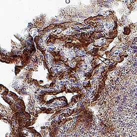
![Immunohistochemistry: OATP2/8 Antibody (MDQ) [NB100-74482] - Immunohistochemical staining of liver transporters in the resected specimens from our case and control cases. Image collected and cropped by CiteAb from the following publication (surgicalcasereports.springeropen.com/articles/10.1186/s40792-016-0216-8), licensed under a CC-BY license.](https://images.novusbio.com/fullsize/OATP2-8-Antibody-MDQ-Immunohistochemistry-NB100-74482-img0003.jpg)
![Western Blot: MRP2 Antibody [NBP1-69023] - Sample Tissue: Mouse Thymus Antibody Dilution: 1.0ug/ml](https://images.novusbio.com/fullsize/MRP2-Antibody-Western-Blot-NBP1-69023-img0004.jpg)
![Western Blot: MRP2 Antibody [NBP1-69023] - Mouse Spleen lysate, concentration 0.2-1 ug/ml.](https://images.novusbio.com/fullsize/MRP2-Antibody-Western-Blot-NBP1-69023-img0003.jpg)
![Immunohistochemistry-Paraffin: UGT Antibody [NBP1-80642] - Staining of human Cerebral cortex shows moderate granular cytoplasmic positivity in neuronal and glial cells.](https://images.novusbio.com/fullsize/UGT-Antibody-Immunohistochemistry-Paraffin-NBP1-80642-img0005.jpg)
![Immunohistochemistry-Paraffin: UGT Antibody [NBP1-80642] - Staining of human rectum shows strong cytoplasmic staining and additional nuclear membrane positivity in glandular cells.](https://images.novusbio.com/fullsize/UGT-Antibody-Immunohistochemistry-Paraffin-NBP1-80642-img0002.jpg)
![Immunohistochemistry-Paraffin: OATP1B3/SLCO1B3/OATP8 Antibody [NBP1-80980] - Staining in human liver and kidney tissues using NBP1-80980 antibody. Corresponding SLCO1B3 RNA-seq data are presented for the same tissues.](https://images.novusbio.com/fullsize/OATP1B3-SLCO1B3-OATP8-Antibody-Immunohistochemistry-Paraffin-NBP1-80980-img0014.jpg)
![Immunocytochemistry/Immunofluorescence: OATP1B3/SLCO1B3/OATP8 Antibody [NBP1-80980] - Staining of human cell line A-431 shows positivity in cytoplasm. Antibody staining is shown in green.](https://images.novusbio.com/fullsize/OATP1B3-SLCO1B3-OATP8-Antibody-Immunocytochemistry-Immunofluorescence-NBP1-80980-img0004.jpg)
![Western Blot: GEN1 Antibody [NBP2-30783] - Analysis in human cell line RH-30.](https://images.novusbio.com/fullsize/GEN1-Antibody-Western-Blot-NBP2-30783-img0004.jpg)
![Immunocytochemistry/Immunofluorescence: GEN1 Antibody [NBP2-30783] - Immunofluorescent staining of human cell line RH-30 shows localization to nucleoplasm.](https://images.novusbio.com/fullsize/GEN1-Antibody-Immunocytochemistry-Immunofluorescence-NBP2-30783-img0006.jpg)


![Immunohistochemistry-Paraffin: MRP3 Antibody [NBP2-37923] - Staining in human stomach and skeletal muscle tissues.. Corresponding ABCC3 RNA-seq data are presented for the same tissues.](https://images.novusbio.com/fullsize/MRP3-Antibody-Immunohistochemistry-Paraffin-NBP2-37923-img0008.jpg)
![Immunocytochemistry/Immunofluorescence: MRP3 Antibody [NBP2-37923] - Immunofluorescent staining of human cell line A549 shows localization to plasma membrane. Antibody staining is shown in green.](https://images.novusbio.com/fullsize/MRP3-Antibody-Immunocytochemistry-Immunofluorescence-NBP2-37923-img0004.jpg)
![Western Blot: Citidine Deaminase Antibody [NBP2-39019] - Lane 1: Marker [kDa] 250, 130, 95, 72, 55, 36, 28, 17, 10. Lane 2: Human cell line RT-4. Lane 3: Human cell line U-251MG. Lane 4: Human Plasma. Lane 5: Human liver tissue](https://images.novusbio.com/fullsize/Citidine-Deaminase-Antibody-Western-Blot-NBP2-39019-img0003.jpg)
![Immunocytochemistry/Immunofluorescence: Citidine Deaminase Antibody [NBP2-39019] - Immunofluorescent staining of human cell line U-2 OS shows localization to nucleoplasm. Antibody staining is shown in green.](https://images.novusbio.com/fullsize/Citidine-Deaminase-Antibody-Immunocytochemistry-Immunofluorescence-NBP2-39019-img0004.jpg)

![Western Blot: Glutamine Synthetase Antibody [NB110-41404] - Analysis of glutamine synthase. 40ug of lysates from mouse (Lanes M), rat (Lane R), pig (Lane P), bovine (Lane B), or human (Lane Hu) retina were probed. A 42 kDa band was identified in lysates from retinas of all species.](https://images.novusbio.com/fullsize/Glutamine-Synthetase-Antibody-Western-Blot-NB110-41404-img0003.jpg)
![Immunocytochemistry/Immunofluorescence: Glutamine Synthetase Antibody [NB110-41404] - Immunofluorescence using NB110-41404. Submitted via verified customer review.](https://images.novusbio.com/fullsize/Glutamine-Synthetase-Antibody-Immunocytochemistry-Immunofluorescence-NB110-41404-img0006.jpg)
![Western Blot: APC Antibody [NB100-91662] - Lane1:Hela cell lysate. Lane2:HEK293T cell lysate. Lane3:Rat testis tissue lysate.](https://images.novusbio.com/fullsize/APC-Antibody-Western-Blot-NB100-91662-img0004.jpg)
![Immunocytochemistry/Immunofluorescence: APC Antibody [NB100-91662] - HeLa cells were fixed for 10 minutes using 10% formalin and then permeabilized for 5 minutes using 1X PBS + 0.05% Triton-X100. The cells were incubated with anti-APC Antibody at 2 ug/ml overnight at 4C and detected with an anti-rabbit Dylight 488 (Green) at a 1:500 dilution. Nuclei were counterstained with DAPI (Blue). Cells were imaged using a 40X objective.](https://images.novusbio.com/fullsize/APC-Antibody-Immunocytochemistry-Immunofluorescence-NB100-91662-img0006.jpg)

![Immunocytochemistry/Immunofluorescence: SQLE Antibody [NBP2-93808] - Analysis of NIH-3T3 cells using SQLE . Blue: DAPI for nuclear staining.](https://images.novusbio.com/fullsize/SQLE-Antibody-Immunocytochemistry-Immunofluorescence-NBP2-93808-img0002.jpg)
![Immunocytochemistry/Immunofluorescence: SQLE Antibody [NBP2-93808] - Analysis of HeLa cells using SQLE . Blue: DAPI for nuclear staining.](https://images.novusbio.com/fullsize/SQLE-Antibody-Immunocytochemistry-Immunofluorescence-NBP2-93808-img0001.jpg)
