Research of Meniere Disease has been linked to Vertigo, Edema, Endolymphatic Hydrops, Sensorineural Hearing Loss (disorder), Labyrinthine Disorder. The study of Meniere Disease has been mentioned in research publications which can be found using our bioinformatics tool below. Researched pathways related to Meniere Disease include Pathogenesis, Reflex, Localization, Transport, Aging. These pathways complement our catalog of research reagents for the study of Meniere Disease including antibodies and ELISA kits against AP, AQUAPORIN 2, ABR, AQP2, AVP.
Top Research Reagents
We have 1276 products for the study of Meniere Disease that can be applied to Chromatin Immunoprecipitation (ChIP), Flow Cytometry, Immunocytochemistry/ Immunofluorescence, Immunohistochemistry, Western Blot from our catalog of antibodies and ELISA kits.
![Immunocytochemistry/Immunofluorescence: Aquaporin-2 Antibody [NB110-74682] - HeLa cells were fixed in 4% paraformaldehyde for 10 minutes and permeabilized in 0.05% Triton X-100 in PBS for 5 minutes. The cells were incubated with anti-Aquaporin-2 Antibody NB110-74682 at 1 ug/ml overnight at 4C and detected with an anti-rabbit Dylight 488 (Green) at a 1:1000 dilution for 60 minutes. Nuclei were counterstained with DAPI (Blue). Cells were imaged using a 100X objective and digitally deconvolved.](https://images.novusbio.com/fullsize/Aquaporin-2-Antibody-Immunocytochemistry-Immunofluorescence-NB110-74682-img0005.jpg)
![Immunocytochemistry/Immunofluorescence: Aquaporin-2 Antibody [NB110-74682] - HepG2 cells were fixed in 4% paraformaldehyde for 10 minutes and permeabilized in 0.05% Triton X-100 in PBS for 5 minutes. The cells were incubated with anti-Aquaporin-2 Antibody NB110-74682 at 1 ug/ml overnight at 4C and detected with an anti-rabbit Dylight 488 (Green) at a 1:1000 dilution for 60 minutes. Nuclei were counterstained with DAPI (Blue). Cells were imaged using a 40X objective.](https://images.novusbio.com/fullsize/Aquaporin-2-Antibody-Immunocytochemistry-Immunofluorescence-NB110-74682-img0006.jpg)
![Western Blot: ABR Antibody [NBP1-30981] - Sample (30ug whole cell lysate) A:A431, 7.5% SDS PAGE, antibody diluted at 1:1000.](https://images.novusbio.com/fullsize/ABR-Antibody-Western-Blot-NBP1-30981-img0010.jpg)
![Immunocytochemistry/Immunofluorescence: ABR Antibody [NBP1-30981] - A431 cells were fixed in 4% paraformaldehyde at RT for 15 min. Green: ABR protein stained by ABR antibody [C3], C-term diluted at 1:500. Blue: Hoechst 33342 staining. Scale bar = 10 um.](https://images.novusbio.com/fullsize/ABR-Antibody-Immunocytochemistry-Immunofluorescence-NBP1-30981-img0009.jpg)
![Western Blot: HMGCL Antibody [NBP1-32767] - rat liver lysate/extract 10 % SDS-PAGE gel, antibody dilution 1:10000.](https://images.novusbio.com/fullsize/HMGCL-Antibody-Western-Blot-NBP1-32767-img0007.jpg)
![Immunocytochemistry/Immunofluorescence: HMGCL Antibody [NBP1-32767] - HeLa cells were fixed in ice-cold MeOH for 5 min. Green: HMGCL protein stained by HMGCL antibody diluted at 1:500. Blue: Hoechst 33342 staining. Scale bar = 10 um.](https://images.novusbio.com/fullsize/HMGCL-Antibody-Immunocytochemistry-Immunofluorescence-NBP1-32767-img0008.jpg)
![Western Blot: HES-1 Antibody (OTI4H1) [NBP1-47791] - HEK293T cells were transfected with the pCMV6-ENTRY control (Left lane) or pCMV6-ENTRY HES1 (Right lane) cDNA for 48 hrs and lysed. Equivalent amounts of cell lysates (5 ug per lane) were separated by SDS-PAGE and immunoblotted with anti-HES1 (1:500).](https://images.novusbio.com/fullsize/HES-1-Antibody-OTI4H1-Western-Blot-NBP1-47791-img0018.jpg)
![Immunocytochemistry/Immunofluorescence: HES-1 Antibody (OTI4H1) [NBP1-47791] - HeLa cells using anti-HES1 mouse monoclonal antibody.](https://images.novusbio.com/fullsize/HES-1-Antibody-OTI4H1-Immunocytochemistry-Immunofluorescence-NBP1-47791-img0010.jpg)
![Western Blot: SRPR alpha Antibody [H00006734-B02P] - Analysis of SRPR expression in human pancreas.](https://images.novusbio.com/fullsize/SRPR-alpha-Antibody-Western-Blot-H00006734-B02P-img0002.jpg)
![Immunocytochemistry/Immunofluorescence: SRPR alpha Antibody [H00006734-B02P] - Analysis of purified antibody to SRPR on HeLa cell. (antibody concentration 10 ug/ml)](https://images.novusbio.com/fullsize/SRPR-alpha-Antibody-Immunocytochemistry-Immunofluorescence-H00006734-B02P-img0001.jpg)


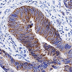
![Immunohistochemistry-Paraffin: LAMC2 Antibody (CL2980) [NBP2-42388] - Staining in human fallopian tube and liver tissues. Corresponding LAMC2 RNA-seq data are presented for the same tissues.](https://images.novusbio.com/fullsize/LAMC2-Antibody-CL2980-Immunohistochemistry-Paraffin-NBP2-42388-img0023.jpg)
![Western Blot: LAMC2 Antibody (CL2980) [NBP2-42388] - Analysis in A-431 cells transfected with control siRNA, target specific siRNA probe #1 and #2, using Anti-LAMC2 antibody. Remaining relative intensity is presented. Loading control: Anti-GAPDH.](https://images.novusbio.com/fullsize/LAMC2-Antibody-CL2980-Western-Blot-NBP2-42388-img0017.jpg)
![Immunohistochemistry-Paraffin: Desmoplakin Antibody [NBP2-48836] - Staining in human skin and skeletal muscle tissues using anti-DSP antibody. Corresponding DSP RNA-seq data are presented for the same tissues.](https://images.novusbio.com/fullsize/Desmoplakin-Antibody-Immunohistochemistry-Paraffin-NBP2-48836-img0005.jpg)
![Immunohistochemistry-Paraffin: Desmoplakin Antibody [NBP2-48836] - Staining of human skeletal muscle shows low expression as expected.](https://images.novusbio.com/fullsize/Desmoplakin-Antibody-Immunohistochemistry-Paraffin-NBP2-48836-img0003.jpg)
![Western Blot: HLA B Antibody (2G7B10) [NBP2-61871] - Analysis using HLA-B mAb against HEK293 (1) and HLA-B (AA: 241-362)-hIgGFc transfected HEK293 (2) cell lysate.](https://images.novusbio.com/fullsize/HLA-B-Antibody-2G7B10-Western-Blot-NBP2-61871-img0008.jpg)
![Immunocytochemistry/Immunofluorescence: HLA B Antibody (2G7B10) [NBP2-61871] - Analysis of Hela cells using HLA-B mouse mAb (green). Blue: DRAQ5 fluorescent DNA dye. Red: Actin filaments have been labeled with Alexa Fluor- 555 phalloidin. Goat anti-Mouse IgG (H+L) DyLight 488 secondary antibody was used.](https://images.novusbio.com/fullsize/HLA-B-Antibody-2G7B10-Immunocytochemistry-Immunofluorescence-NBP2-61871-img0004.jpg)
![Immunocytochemistry/ Immunofluorescence: LIPC Antibody - BSA Free [NBP3-04922] - Immunofluorescence analysis of A549 cells using LIPC antibody (A13508). Blue: DAPI for nuclear staining.](https://images.novusbio.com/fullsize/nbp3-04922_rabbit-polyclonal-lipc-antibody-5220251931326.jpg)
![Western Blot: PTCRA Antibody [NBP3-09949] - Western blot analysis of PTCRA in HT1080 Whole Cell lysates. Antibody dilution at 1.0ug/ml](https://images.novusbio.com/fullsize/PTCRA-Antibody-Western-Blot-NBP3-09949-img0001.jpg)
