| Submit your blog on Nystagmus to be featured! |
| Submit your event on Nystagmus to be featured! |
Mouse Monoclonal
Applications IHC
|
|
Rabbit Polyclonal
Species Human, Rat
Applications WB, IHC
|
|
![Western Blot: SRPR alpha Antibody [H00006734-B02P] - Analysis of SRPR expression in human pancreas.](https://images.novusbio.com/fullsize/SRPR-alpha-Antibody-Western-Blot-H00006734-B02P-img0002.jpg)
![Immunocytochemistry/Immunofluorescence: SRPR alpha Antibody [H00006734-B02P] - Analysis of purified antibody to SRPR on HeLa cell. (antibody concentration 10 ug/ml)](https://images.novusbio.com/fullsize/SRPR-alpha-Antibody-Immunocytochemistry-Immunofluorescence-H00006734-B02P-img0001.jpg)
Mouse Polyclonal
Species Human
Applications WB, ICC/IF
| 1 Publication |
|
![Immunohistochemistry-Paraffin: CRAT Antibody [NBP1-86616] - Analysis in human testis and lymph node tissues. Corresponding CRAT RNA-seq data are presented for the same tissues.](https://images.novusbio.com/fullsize/CRAT-Antibody-Immunohistochemistry-Paraffin-NBP1-86616-img0014.jpg)
![Western Blot: CRAT Antibody [NBP1-86616] - Analysis using Anti-CRAT antibody NBP1-86616 (A) shows similar pattern to independent antibody NBP1-86615 (B).](https://images.novusbio.com/fullsize/CRAT-Antibody-Western-Blot-NBP1-86616-img0011.jpg)
Rabbit Polyclonal
Species Human
Applications WB, IHC, IHC-P
| 2 Publications |
|
![Immunohistochemistry-Paraffin: Myelin PLP Antibody [NBP1-87781] - Staining in human cerebral cortex and pancreas tissues using anti-PLP1 antibody. Corresponding PLP1 RNA-seq data are presented for the same tissues.](https://images.novusbio.com/fullsize/Myelin-PLP-Antibody-Immunohistochemistry-Paraffin-NBP1-87781-img0012.jpg)
![Immunohistochemistry-Paraffin: Myelin PLP Antibody [NBP1-87781] - Border of a demyelinated lesion. Image submitted by a verified customer review.](https://images.novusbio.com/fullsize/Myelin-PLP-Antibody-Immunohistochemistry-Paraffin-NBP1-87781-img0013.jpg)
Rabbit Polyclonal
Species Human, Mouse
Applications WB, IHC, IHC-P
| 1 Review 5 Publications |
|
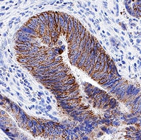
![Immunohistochemistry-Paraffin: FRMD7 Antibody [NBP1-93762] - Staining of human stomach shows strong cytoplasmic positivity in glandular cells.](https://images.novusbio.com/fullsize/FRMD7-Antibody-Immunohistochemistry-Paraffin-NBP1-93762-img0004.jpg)
Rabbit Polyclonal
Species Human
Applications IHC, IHC-P
| 4 Publications |
|
![Immunohistochemistry-Paraffin: OA1 Antibody [NBP2-14066] - Staining in human skin and skeletal muscle tissues . Corresponding GPR143 RNA-seq data are presented for the same tissues.](https://images.novusbio.com/fullsize/OA1-Antibody-Immunohistochemistry-Paraffin-NBP2-14066-img0005.jpg)
![Immunohistochemistry-Paraffin: OA1 Antibody [NBP2-14066] - Staining of human skin shows moderate to strong cytoplasmic positivity in melanocytes.](https://images.novusbio.com/fullsize/OA1-Antibody-Immunohistochemistry-Paraffin-NBP2-14066-img0004.jpg)
Rabbit Polyclonal
Species Human
Applications IHC, IHC-P
|
|


Goat Polyclonal
Species Human, Mouse, Rat
Applications WB, Simple Western, ICC
| 3 Reviews 15 Publications |
|


Sheep Polyclonal
Species Human
Applications WB, Simple Western, ICC
| 15 Publications |
|
![Western Blot: hnRNP C1 + C2 Antibody (CL2593) [NBP2-36776] - Analysis in U-251MG cells transfected with control siRNA, target specific siRNA probe #1 and #2, using Anti-HNRNPC antibody. Remaining relative intensity is presented. Loading control: Anti-PPIB.](https://images.novusbio.com/fullsize/hnRNP-C1-+-C2-Antibody-CL2593-Western-Blot-NBP2-36776-img0015.jpg)
![Immunocytochemistry/Immunofluorescence: hnRNP C1 + C2 Antibody (CL2593) [NBP2-36776] - Staining in U2OS cell line with Anti-HNRNPC monoclonal antibody, showing distinct nuclear (without nucleoli) staining in green. Microtubule- and nuclear probes are visualized in red and blue respectively (where available). Antibody staining is shown in green.](https://images.novusbio.com/fullsize/hnRNP-C1-+-C2-Antibody-CL2593-Immunocytochemistry-Immunofluorescence-NBP2-36776-img0014.jpg)
Mouse Monoclonal
Species Human
Applications WB, ICC/IF, IHC
|
|


Species Human
Applications BA
| 21 Publications |
|

Species Human
Applications BA
| 3 Publications |
|
![Immunohistochemistry-Paraffin: LAMC2 Antibody (CL2980) [NBP2-42388] - Staining in human fallopian tube and liver tissues. Corresponding LAMC2 RNA-seq data are presented for the same tissues.](https://images.novusbio.com/fullsize/LAMC2-Antibody-CL2980-Immunohistochemistry-Paraffin-NBP2-42388-img0023.jpg)
![Western Blot: LAMC2 Antibody (CL2980) [NBP2-42388] - Analysis in A-431 cells transfected with control siRNA, target specific siRNA probe #1 and #2, using Anti-LAMC2 antibody. Remaining relative intensity is presented. Loading control: Anti-GAPDH.](https://images.novusbio.com/fullsize/LAMC2-Antibody-CL2980-Western-Blot-NBP2-42388-img0017.jpg)
Mouse Monoclonal
Species Human
Applications WB, ICC/IF, IHC
| 4 Publications |
|
![Immunohistochemistry-Paraffin: Desmoplakin Antibody [NBP2-48836] - Staining in human skin and skeletal muscle tissues using anti-DSP antibody. Corresponding DSP RNA-seq data are presented for the same tissues.](https://images.novusbio.com/fullsize/Desmoplakin-Antibody-Immunohistochemistry-Paraffin-NBP2-48836-img0005.jpg)
![Immunohistochemistry-Paraffin: Desmoplakin Antibody [NBP2-48836] - Staining of human skeletal muscle shows low expression as expected.](https://images.novusbio.com/fullsize/Desmoplakin-Antibody-Immunohistochemistry-Paraffin-NBP2-48836-img0003.jpg)
Rabbit Polyclonal
Species Human, Canine
Applications ICC/IF, IHC, IHC-P
| 2 Publications |
|
![Western Blot: GLYAT Antibody [NBP2-58917] - Western blot analysis in human cell line RT-4, human cell line U-251 MG, human plasma and human liver tissue.](https://images.novusbio.com/fullsize/GLYAT-Antibody-Western-Blot-NBP2-58917-img0002.jpg)
![Immunohistochemistry-Paraffin: GLYAT Antibody [NBP2-58917] - Staining of human skeletal muscle.](https://images.novusbio.com/fullsize/GLYAT-Antibody-Immunohistochemistry-Paraffin-NBP2-58917-img0010.jpg)
Rabbit Polyclonal
Species Human
Applications WB, IHC, IHC-P
|
|
![Western Blot: ERG Antibody [NBP2-60655] - Total protein from Human THP-1, Jurkat and K562 cells was separated on a 7.5% gel by SDS-PAGE, transferred to PVDF membrane and blocked in 5% non-fat milk in TBST. The membrane was probed with 2.0 ug/ml anti-ERG in blocking buffer and detected with an anti-rabbit HRP secondary antibody using chemiluminescence.](https://images.novusbio.com/fullsize/ERG-Antibody-Western-Blot-NBP2-60655-img0002.jpg)
![Immunocytochemistry/Immunofluorescence: ERG Antibody [NBP2-60655] - HeLa cells were fixed for 10 minutes using 10% formalin and then permeabilized for 5 minutes using 1X TBS + 0.5% Triton-X100. The cells were incubated with anti-ERG at 5 ug/ml overnight at 4C and detected with an anti-rabbit Dylight 488 (Green) at a 1:500 dilution. Alpha tubulin (DM1A) NB100-690 was used as a co-stain at a 1:1000 dilution and detected with an anti-mouse Dylight 550 (Red) at a 1:500 dilution. Nuclei were counterstained with DAPI (Blue). Cells were imaged using a 40X objective.](https://images.novusbio.com/fullsize/ERG-Antibody-Immunocytochemistry-Immunofluorescence-NBP2-60655-img0001.jpg)
Rabbit Polyclonal
Species Human
Applications WB, ICC/IF, IHC
|
|
![Western Blot: Tyrosinase Antibody (JA52-11) [NBP2-67232] - Western blot analysis of Tyrosinase on B16F1 cell lysates. Proteins were transferred to a PVDF membrane and blocked with 5% BSA in PBS for 1 hour at room temperature. The primary antibody (1/500) was used in 5% BSA at room temperature for 2 hours. Goat Anti-Rabbit IgG - HRP Secondary Antibody (HA1001) at 1:200,000 dilution was used for 1 hour at room temperature.](https://images.novusbio.com/fullsize/Tyrosinase-Antibody-JA52-11-Western-Blot-NBP2-67232-img0006.jpg)
![Immunocytochemistry/Immunofluorescence: Tyrosinase Antibody (JA52-11) [NBP2-67232] - Staining Tyrosinase in B16F1 cells (red). The nuclear counter stain is DAPI (blue). Cells were fixed in paraformaldehyde, permeabilised with 0.25% Triton X100/PBS.](https://images.novusbio.com/fullsize/Tyrosinase-Antibody-JA52-11-Immunocytochemistry-Immunofluorescence-NBP2-67232-img0005.jpg)
Rabbit Monoclonal
Species Human, Mouse
Applications WB, ICC/IF, IHC
|
|
![Western Blot: Kv11.1 Antibody [NBP3-03109] - Analysis of extracts of various cell lines, using Kv11.1 antibody at 1:500 dilution. Secondary antibody: HRP Goat Anti-Rabbit IgG (H+L) at 1:10000 dilution. Lysates/proteins: 25ug per lane. Blocking buffer: 3% nonfat dry milk in TBST. Detection: ECL Enhan](https://images.novusbio.com/fullsize/Kv11.1-Antibody-Western-Blot-NBP3-03109-img0004.jpg)
![Immunohistochemistry-Paraffin: Kv11.1 Antibody [NBP3-03109] - Human lung cancer using KCNH2 antibody at dilution of 1:100 (40x lens).Perform microwave antigen retrieval with 10 mM PBS buffer pH 7.2 before commencing with IHC staining protocol.](https://images.novusbio.com/fullsize/Kv11.1-Antibody-Immunohistochemistry-Paraffin-NBP3-03109-img0003.jpg)
Rabbit Polyclonal
Species Human, Mouse
Applications WB, IHC, IHC-P
|
|
