| Submit your blog on Limb Deformities, Congenital to be featured! |
| Submit your event on Limb Deformities, Congenital to be featured! |
![Immunocytochemistry/Immunofluorescence: HOXA13 Antibody [NBP3-17383] - Staining of human cell line RT4 shows localization to nucleoplasm & mitotic chromosome.](https://images.novusbio.com/fullsize/HOXA13-Antibody-Immunocytochemistry-Immunofluorescence-NBP3-17383-img0001.jpg)
![Immunohistochemistry-Paraffin: HOXA13 Antibody [NBP3-17383] - Staining of mouse embryonic limb using HOXA13 antibody. Image from verified customer review.](https://images.novusbio.com/fullsize/nbp3-17383_rabbit-hoxa13-pab-immunohistochemistry-paraffin-284202314556.jpg)
Rabbit Polyclonal
Species Human
Applications ICC/IF, IHC-P
|
|
![Western Blot: UVRAG Antibody [NBP1-18885] - Beclin 1 is acetylated at lysines 430 and 437. TSA and NAM increase the binding of Beclin 1 to Rubicon. Immunoprecipitation of indicated Beclin 1-binding partners with ectopically expressed Flag-Beclin 1 in HEK293T cells treated with TSA and NAM. Image collected and cropped by CiteAb from the following publication (https://www.nature.com/articles/ncomms8215), licensed under a CC-BY license.](https://images.novusbio.com/fullsize/UVRAG-Antibody-Western-Blot-NBP1-18885-img0005.jpg)
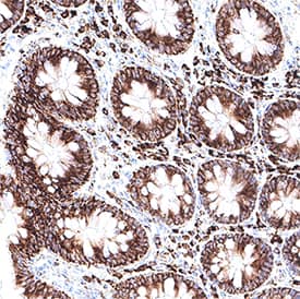
![Immunohistochemistry-Paraffin: UVRAG Antibody [NBP1-18885] - Section of human colon carcinoma. Antibody: Affinity purified rabbit anti- UVRAG used at a dilution of 1:200 (1ug/ml). Detection: DAB](https://images.novusbio.com/fullsize/UVRAG-Antibody-Immunohistochemistry-Paraffin-NBP1-18885-img0004.jpg)
Rabbit Polyclonal
Species Human
Applications WB, ICC/IF, IHC
| 4 Publications |
|
Mouse Monoclonal
Species Human
Applications WB, Simple Western, IHC
|
|
![Immunocytochemistry/Immunofluorescence: HOXD13 Antibody [NBP1-52149] - Immunofluorescence analysis of paraformaldehyde fixed U2OS cells, permeabilized with 0.15% Triton. Primary incubation 1hr (10 ug/mL) followed by Alexa Fluor 488 secondary antibody (2 ug/mL), showing nuclear staining. The nuclear stain is DAPI (blue). Negative control: Unimmunized goat IgG (10 ug/mL) followed by Alexa Fluor 488 secondary antibody (2 ug/mL).](https://images.novusbio.com/fullsize/HOXD13-Antibody-Immunocytochemistry-Immunofluorescence-NBP1-52149-img0003.jpg)
![Immunohistochemistry-Paraffin: HOXD13 Antibody [NBP1-52149] - Staining of Human Prostate. Antibody at 3.8 ug/mL. Steamed antigen retrieval with citrate buffer pH 6, AP-staining.](https://images.novusbio.com/fullsize/HOXD13-Antibody-Immunohistochemistry-Paraffin-NBP1-52149-img0001.jpg)
Goat Polyclonal
Species Human
Applications ICC/IF, IHC, IHC-P
| 1 Review 1 Publication |
|
![Immunohistochemistry-Paraffin: TBX5 Antibody [NBP1-83237] - Staining of human placenta shows moderate nuclear positivity in Hofbauer cells, as well as weaker positivity in trophoblastic cells.](https://images.novusbio.com/fullsize/TBX5-Antibody-Immunohistochemistry-Paraffin-NBP1-83237-img0016.jpg)
![Immunohistochemistry-Paraffin: TBX5 Antibody [NBP1-83237] - Staining of human heart muscle shows strong nuclear positivity in cardiomyocytes.](https://images.novusbio.com/fullsize/TBX5-Antibody-Immunohistochemistry-Paraffin-NBP1-83237-img0013.jpg)
Rabbit Polyclonal
Species Human, Mouse
Applications WB, ICC/IF, IHC
| 4 Publications |
|
![Western Blot: SHFM3 Antibody [NBP2-14013] - Analysis in human cell line MCF-7.](https://images.novusbio.com/fullsize/SHFM3-Antibody-Western-Blot-NBP2-14013-img0010.jpg)
![Immunocytochemistry/Immunofluorescence: SHFM3 Antibody [NBP2-14013] - Immunofluorescent staining of human cell line U-251 MG shows localization to the Golgi apparatus.](https://images.novusbio.com/fullsize/SHFM3-Antibody-Immunocytochemistry-Immunofluorescence-NBP2-14013-img0005.jpg)
Rabbit Polyclonal
Species Human
Applications WB, ICC/IF, IHC
|
|


Goat Polyclonal
Species Human
Applications WB, Simple Western
| 15 Publications |
|


Goat Polyclonal
Species Human, Mouse
Applications WB, ChIP, ICC
| 3 Reviews 102 Publications |
|

Goat Polyclonal
Species Mouse
Applications WB, IHC
| 3 Publications |
|


Goat Polyclonal
Species Mouse
Applications WB, IHC
| 6 Publications |
|


Goat Polyclonal
Species Human
Applications WB, IHC, ICC
| 1 Review 24 Publications |
|


Species Mouse
Applications BA
| 70 Publications |
|


Species Human, Mouse
Applications BA
| 1 Review 63 Publications |
|

Species Human, Mouse, Rat
Applications BA
| 191 Publications |
|


Species Human
Applications BA, BA
| 530 Publications |
|


Species Human
Applications BA
| 76 Publications |
|
![Western Blot: RPE65 Antibody (401.8B11.3D9) - BSA Free [NB100-355] - Subcellular localization of BEST1 and surface Ca2+-dependent Cl- current in patient-derived iPSC-RPEs. Western blots show similar BEST1 expression levels in WT and patient-derived iPSC-RPEs. Each sample was from one cell lysis (BEST1 and beta-actin, RPE65 and CRALBP were on two gels, respectively). Image collected and cropped by CiteAb from the following publication (https://elifesciences.org/articles/29914), licensed under a CC-BY license.](https://images.novusbio.com/fullsize/RPE65-Antibody-401-8B11-3D9-BSA-Free-Western-Blot-NB100-355-img0013.jpg)
![Immunocytochemistry/Immunofluorescence: RPE65 Antibody (401.8B11.3D9) - BSA Free [NB100-355] - Expression of eye-specific markers in the induced eye-like structures induced from lignin-added ES cells. (a) Higher-magnification image of the RPE like structure induced from ESCs after 12-day culture. (b) Immunostaining of eye- like structures. Eye- like structures induced from ESCs after 12-day culture were stained with antibodies against RPE65 (red) and nuclei were stained with DAPI solution (blue). Scale bar: a = 50 um, b,d = 200 um, c,e = 100 um. PLoS One. 2013 Jun 21;8(6):e66376. doi: 10.1371/journal.pone.0066376.](https://images.novusbio.com/fullsize/RPE65-Antibody-401-8B11-3D9-BSA-Free-Immunocytochemistry-Immunofluorescence-NB100-355-img0014.jpg)
Mouse Monoclonal
Species Human, Mouse, Rat
Applications WB, Simple Western, Flow
| 7 Reviews 102 Publications |
|
