| Submit your blog on Andersen Syndrome to be featured! |
| Submit your event on Andersen Syndrome to be featured! |
![Western Blot: KCNE1 Antibody (5B12) [H00003753-M01] - Analysis of KCNE1 over-expressed 293 cell line, cotransfected with KCNE1 Validated Chimera RNAi ( Cat # H00003753-R01V ) (Lane 2) or non-transfected control (Lane 1). Blot probed with KCNE1 monoclonal antibody (M01), clone 5B12 (Cat # H00003753-M01 ). GAPDH ( 36.1 kDa ) used as specificity and loading control.](https://images.novusbio.com/fullsize/KCNE1-Antibody-5B12-Western-Blot-H00003753-M01-img0004.jpg)
![Western Blot: KCNE1 Antibody (5B12) [H00003753-M01] - analysis of KCNE1 expression in transfected 293T cell line by KCNE1 monoclonal antibody (M01), clone 5B12. Lane 1: KCNE1 transfected lysate (14.7 KDa). Lane 2: Non-transfected lysate.](https://images.novusbio.com/fullsize/KCNE1-Antibody-5B12-Western-Blot-H00003753-M01-img0001.jpg)
Mouse Monoclonal
Species Human, Rat, Canine
Applications WB, ELISA, ICC/IF
| 4 Publications |
|
![Immunohistochemistry-Frozen: ABCA4 Antibody (3F4) [NBP1-30032] - IHC-Fr of normal feline retina with antibodies ABCA4 3F4 (red) and HCAR (green). ABCA4 antibody (1:1000) labels outer segments of photoreceptors and HCAR antibody labels cones. IHC-Fr image submitted by a verified customer review.](https://images.novusbio.com/fullsize/ABCA4-Antibody-3F4-Immunohistochemistry-Frozen-NBP1-30032-img0006.jpg)
![Immunohistochemistry: ABCA4 Antibody (3F4) [NBP1-30032] - staining of adult mouse retina showing specific immunolabeling of the ABCA4 protein. Photo courtesy of Mary Raven, University of California, Santa Barbara, CA.](https://images.novusbio.com/fullsize/ABCA4-Antibody-3F4-Immunohistochemistry-NBP1-30032-img0001.jpg)
Mouse Monoclonal
Species Human, Mouse, Bovine
Applications WB, ICC/IF, IHC
| 1 Review 5 Publications |
|
Mouse Monoclonal
Species Human
Applications WB, Flow, ICC/IF
|
|
![Western Blot: Kv7.1 Antibody (S37A/10) [NBP2-12897] - Western Blot analysis of Human Cell lysates showing detection of Kv7.1 protein using Mouse Anti-Kv7.1 Monoclonal Antibody, Clone N37A/10 (NBP2-12897). Load: 15 ug. Block: 1.5% BSA for 30 minutes at RT. Primary Antibody: Mouse Anti-Kv7.1 Monoclonal Antibody (NBP2-12897) at 1:1000 for 2 hours at RT. Secondary Antibody: Sheep Anti-Mouse IgG: HRP for 1 hour at RT.](https://images.novusbio.com/fullsize/Kv7-1-Antibody-S37A-10-Western-Blot-NBP2-12897-img0010.jpg)
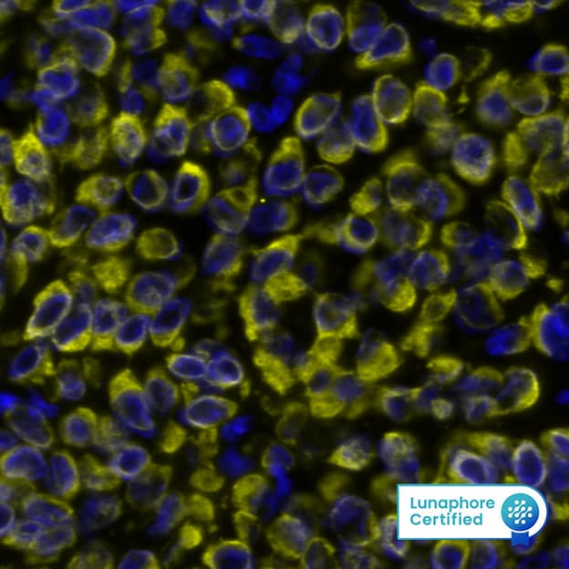
![Immunocytochemistry/Immunofluorescence: Kv7.1 Antibody (S37A/10) [NBP2-12897] - Immunocytochemistry/Immunofluorescence analysis using Mouse Anti-Kv7.1 Monoclonal Antibody, Clone N37A/10 (NBP2-12897). Tissue: Neuroblastoma cells (SH-SY5Y). Species: Human. Fixation: 4% PFA for 15 min. Primary Antibody: Mouse Anti-Kv7.1 Monoclonal Antibody (NBP2-12897) at 1:100 for overnight at 4C with slow rocking. Secondary Antibody: AlexaFluor 488 at 1:1000 for 1 hour at RT. Counterstain: Phalloidin-iFluor 647 (red) F-Actin stain; Hoechst (blue) nuclear stain at 1:800, 1.6mM for 20 min at RT. (A) Hoechst (blue) nuclear stain. (B) Phalloidin-iFluor 647 (red) F-Actin stain. (C) Kv7.1 Antibody (D) Composite.](https://images.novusbio.com/fullsize/Kv7-1-Antibody-S37A-10-Immunocytochemistry-Immunofluorescence-NBP2-12897-img0013.jpg)
Mouse Monoclonal
Species Human, Mouse, Rat
Applications WB, ICC/IF, IHC
|
|
Mouse Monoclonal
Species Human, Mouse, Rat
Applications WB, Flow, ICC/IF
| 1 Review 71 Publications |
|
![Immunohistochemistry-Paraffin: KCNN4 Antibody [NBP2-33694] - Staining of human testis shows no positivity in cells in seminiferous ducts as expected.](https://images.novusbio.com/fullsize/KCNN4-Antibody-Immunohistochemistry-Paraffin-NBP2-33694-img0005.jpg)
![Immunohistochemistry-Paraffin: KCNN4 Antibody [NBP2-33694] - Staining of human colon shows strong cytoplasmic/ membranous positivity in smooth muscle cells.](https://images.novusbio.com/fullsize/KCNN4-Antibody-Immunohistochemistry-Paraffin-NBP2-33694-img0002.jpg)
Rabbit Polyclonal
Species Human
Applications ICC/IF, IHC, IHC-P
| 1 Publication |
|
![Western Blot: Ankyrin Brain Antibody [NBP2-33863] - Lane 1: Marker [kDa] 230, 130, 95, 72, 56, 36, 28, 17, 11. Lane 2: Human cell line RT-4. Lane 3: Human cell line U-251MG sp. Lane 4: Human plasma (IgG/HSA depleted). Lane 5: Human liver tissue. Lane 6: Human tonsil tissue](https://images.novusbio.com/fullsize/Ankyrin-Brain-Antibody-Western-Blot-NBP2-33863-img0003.jpg)
![Immunocytochemistry/Immunofluorescence: Ankyrin Brain Antibody [NBP2-33863] - Staining of human cell line RH-30 shows localization to plasma membrane. Antibody staining is shown in green.](https://images.novusbio.com/fullsize/Ankyrin-Brain-Antibody-Immunocytochemistry-Immunofluorescence-NBP2-33863-img0008.jpg)
Rabbit Polyclonal
Species Human
Applications WB, ICC/IF, IHC
|
|


Goat Polyclonal
Species Human
Applications WB, Simple Western, ChIP
| 2 Publications |
|
Mouse Monoclonal
Species Human
Applications Flow, Block, CyTOF-reported
| 5 Publications |
|
![Immunohistochemistry-Paraffin: MiRP1 Antibody [NBP2-38146] - Staining of human testis.](https://images.novusbio.com/fullsize/MiRP1-Antibody-Immunohistochemistry-Paraffin-NBP2-38146-img0006.jpg)
![Immunohistochemistry: MiRP1 Antibody [NBP2-38146] - Staining of human stomach shows strong cytoplasmic positivity in subset of glandular cells.](https://images.novusbio.com/fullsize/MiRP1-Antibody-Immunohistochemistry-NBP2-38146-img0001.jpg)
Rabbit Polyclonal
Species Human
Applications IHC, IHC-P
|
|
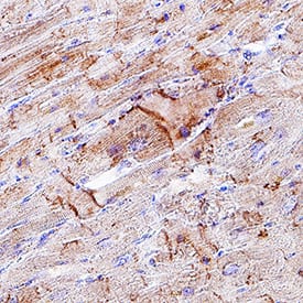
![Immunohistochemistry-Paraffin: CACNA1S Antibody (1A) [NB300-542] - Immunohistochemistry was performed on normal biopsies of deparaffinized human skeletal muscle tissue.](https://images.novusbio.com/fullsize/CACNA1S-Antibody-1A-Immunohistochemistry-Paraffin-NB300-542-img0002.jpg)
![Flow Cytometry: CACNA1S Antibody (1A) [NB300-542] - Flow cytometry analysis of Dihydropyridine Receptor alpha-1 in U251 cells (green) compared to an isotype control (blue). Cells were harvested, adjusted to a concentration of 1-5x10^6 cells/ml, fixed with 2% paraformaldehyde and washed with PBS.](https://images.novusbio.com/fullsize/CACNA1S-Antibody-1A-Flow-Cytometry-NB300-542-img0003.jpg)
Mouse Monoclonal
Species Human, Mouse, Rat
Applications WB, Flow, IHC
| 2 Publications |
|

Rabbit Monoclonal
Species Human, Mouse
Applications WB, IHC, ICC
| 2 Publications |
|
![Western Blot: Kir2.2 Antibody [NBP2-87693] - Host: Rabbit. Target Name: KCNJ12. Sample Type: Human Adult Placenta. Antibody Dilution: 1.0ug/ml](https://images.novusbio.com/fullsize/Kir2.2-Antibody-Western-Blot-NBP2-87693-img0009.jpg)
![Immunohistochemistry-Paraffin: Kir2.2 Antibody [NBP2-87693] - Rabbit Anti-KCNJ12 antibody. Formalin Fixed Paraffin Embedded Tissue: Human Adult Skeletal muscle. Observed Staining: Cytoplasm in hepatocytes. Primary Antibody Concentration: 1:600. Secondary Antibody: Donkey anti-Rabbit-Cy3. Se](https://images.novusbio.com/fullsize/Kir2.2-Antibody-Immunohistochemistry-Paraffin-NBP2-87693-img0001.jpg)
Rabbit Polyclonal
Species Human
Applications WB, IHC-P
|
|
![Western Blot: KIR2.3 Antibody [NBP3-03005] - Analysis of extracts of various cell lines, using KIR2.3 antibody at 1:1000 dilution. Secondary antibody: HRP Goat Anti-Rabbit IgG (H+L) at 1:10000 dilution. Lysates/proteins: 25ug per lane. Blocking buffer: 3% nonfat dry milk in TBST. Detection: ECL Enhanced Kit](https://images.novusbio.com/fullsize/KIR2.3-Antibody-Western-Blot-NBP3-03005-img0002.jpg)
![Immunohistochemistry-Paraffin: KIR2.3 Antibody [NBP3-03005] - Paraffin-embedded rat brain using KIR2.3 antibody at dilution of 1:100 (40x lens).](https://images.novusbio.com/fullsize/KIR2.3-Antibody-Immunohistochemistry-Paraffin-NBP3-03005-img0001.jpg)
Rabbit Polyclonal
Species Human, Mouse, Rat
Applications WB, IHC, IHC-P
|
|
![Western Blot: Kv11.1 Antibody [NBP3-03109] - Analysis of extracts of various cell lines, using Kv11.1 antibody at 1:500 dilution. Secondary antibody: HRP Goat Anti-Rabbit IgG (H+L) at 1:10000 dilution. Lysates/proteins: 25ug per lane. Blocking buffer: 3% nonfat dry milk in TBST. Detection: ECL Enhan](https://images.novusbio.com/fullsize/Kv11.1-Antibody-Western-Blot-NBP3-03109-img0004.jpg)
![Immunohistochemistry-Paraffin: Kv11.1 Antibody [NBP3-03109] - Human lung cancer using KCNH2 antibody at dilution of 1:100 (40x lens).Perform microwave antigen retrieval with 10 mM PBS buffer pH 7.2 before commencing with IHC staining protocol.](https://images.novusbio.com/fullsize/Kv11.1-Antibody-Immunohistochemistry-Paraffin-NBP3-03109-img0003.jpg)
Rabbit Polyclonal
Species Human, Mouse
Applications WB, IHC, IHC-P
|
|
![Immunohistochemistry-Paraffin: GLUT10 Antibody [NBP3-12262] - IHC of NBP3-12262 with mouse brain. Short perfusion followed by 1 hour post-fixation and immersion in sucrose. 1:100 antibody dilution in DiluObuffer.](https://images.novusbio.com/fullsize/GLUT10-Antibody-Immunohistochemistry-Paraffin-NBP3-12262-img0001.jpg)
Rabbit Polyclonal
Species Human, Mouse, Rat
Applications WB, ELISA, ICC/IF
|
|
