| Submit your blog on Acute Respiratory Infections to be featured! |
| Submit your event on Acute Respiratory Infections to be featured! |
![Western Blot: GPHA2 Antibody [NBP3-17255] - Lane 1: Marker [kDa] 250, 130, 95, 72, 55, 36, 28, 17, 10; Lane 2: RT4; Lane 3: U-251 MG; Lane 4: Human Plasma; Lane 5: Liver; Lane 6: Tonsil](https://images.novusbio.com/fullsize/GPHA2-Antibody-Western-Blot-NBP3-17255-img0002.jpg)
![Immunocytochemistry/Immunofluorescence: GPHA2 Antibody [NBP3-17255] - Staining of human cell line Hep G2 shows localization to vesicles.](https://images.novusbio.com/fullsize/GPHA2-Antibody-Immunocytochemistry-Immunofluorescence-NBP3-17255-img0001.jpg)
Rabbit Polyclonal
Species Human
Applications WB, ICC/IF
|
|
![N/A C-Reactive Protein/CRP [HRP]](https://images.novusbio.com/fullsize2/dcrp00b_human-c-reactive-protein-crp-quantikine-elisa-kit-14122022142042.jpg)
![N/A C-Reactive Protein/CRP [HRP]](https://images.novusbio.com/fullsize2/dcrp00b_human-c-reactive-protein-crp-quantikine-elisa-kit-14122022142115.jpg)
Species Human
Applications ELISA
| 150 Publications |
|
![N/A IL-6 [HRP]](https://images.novusbio.com/fullsize/d6050b_human-il-6-quantikine-elisa-kit-2762023115954.png)
![N/A IL-6 [HRP]](https://images.novusbio.com/fullsize/d6050b_human-il-6-quantikine-elisa-kit-276202312352.jpg)
Species Human
| 785 Publications |
|
Rabbit Polyclonal
Species Human
Applications WB, ICC/IF, IHC
|
|
![Western Blot: MS4A1/CD20 Antibody (AISB12) [NBP1-43435] - Immunoblot of Balb/c thymus (lane 1) and A20 (lane 2) cell lysates with Anti-Mouse CD20 Purified.](https://images.novusbio.com/fullsize/MS4A1-CD20-Antibody-AISB12-Western-Blot-NBP1-43435-img0002.jpg)
![Flow Cytometry: MS4A1/CD20 Antibody (AISB12) [NBP1-43435] - Staining of BALB/c splenocytes with Anti-Mouse CD19 Alexa Fluor (R) 647 and 1.0 micrograms conjugated anti-Mouse CD20 Purified followed by Anti-Rat IgG PE. Quadrant lines represent Rat IgG2a isotype control staining levels and cells in the lymphocyte gate were used for analysis.](https://images.novusbio.com/fullsize/MS4A1-CD20-Antibody-AISB12-Flow-Cytometry-NBP1-43435-img0004.jpg)
Rat Monoclonal
Species Human, Mouse
Applications WB, Flow
| 3 Publications |
|
![Western Blot: ATP6V0A2 Antibody [NBP1-59069] - HeLa cells, concentration 3.3 ug/ml.](https://images.novusbio.com/fullsize/ATP6V0A2-Antibody-Western-Blot-NBP1-59069-img0002.jpg)
![Immunohistochemistry: ATP6V0A2 Antibody [NBP1-59069] - Human kidney lysate tissue at an antibody concentration of 5.0 ug/ml using anti-ATP6V0A2 antibody](https://images.novusbio.com/fullsize/ATP6V0A2-Antibody-Immunohistochemistry-NBP1-59069-img0008.jpg)
Rabbit Polyclonal
Species Human
Applications WB, ICC/IF, IHC
|
|
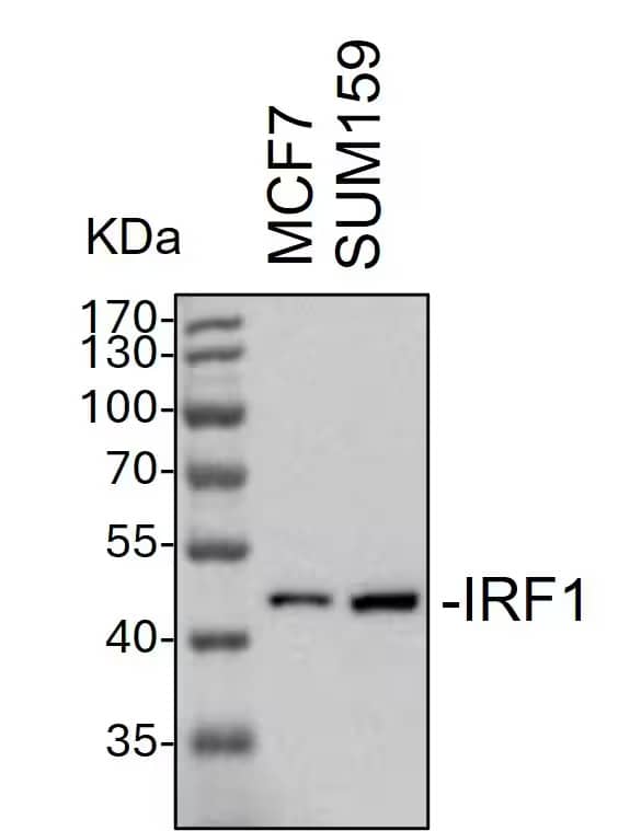
![Western Blot: URI Antibody [NBP1-79413] - MCF7, Antibody Dilution: 1.0 ug/ml There is BioGPS gene expression data showing that URI1 is expressed in MCF7.](https://images.novusbio.com/fullsize/URI-Antibody-Western-Blot-NBP1-79413-img0006.jpg)
![Western Blot: URI Antibody [NBP1-79413] - Human Muscle lysate, concentration 0.2-1 ug/ml.](https://images.novusbio.com/fullsize/URI-Antibody-Western-Blot-NBP1-79413-img0002.jpg)
Rabbit Polyclonal
Species Human
Applications WB
|
|
![Immunohistochemistry-Paraffin: Ornithine Carbamoyltransferase Antibody [NBP1-87408] - Staining in human liver and kidney tissues . Corresponding OTC RNA-seq data are presented for the same tissues.](https://images.novusbio.com/fullsize/Ornithine-Carbamoyltransferase-Antibody-Immunohistochemistry-Paraffin-NBP1-87408-img0020.jpg)
![Immunohistochemistry-Paraffin: Ornithine Carbamoyltransferase Antibody [NBP1-87408] - Staining of human cerebral cortex, kidney, liver and small intestine using Anti-OTC antibody NBP1-87408 (A) shows similar protein distribution across tissues to independent antibody NBP1-88121 (B).](https://images.novusbio.com/fullsize/Ornithine-Carbamoyltransferase-Antibody-Immunohistochemistry-Paraffin-NBP1-87408-img0027.jpg)
Rabbit Polyclonal
Species Human, Mouse
Applications WB, Simple Western, ICC/IF
| 5 Publications |
|
![Immunohistochemistry-Paraffin: BBS9 Antibody [NBP1-88927] - Staining of human cerebellum shows strong cytoplasmic positivity in purkinje cells.](https://images.novusbio.com/fullsize/BBS9-Antibody-Immunohistochemistry-Paraffin-NBP1-88927-img0002.jpg)
Rabbit Polyclonal
Species Human, Mouse
Applications WB, ICC/IF, IHC
| 3 Publications |
|
![Western Blot: NDUFB6 Antibody [NBP1-92172] - Lane 1: Marker [kDa] 250, 130, 95, 72, 55, 36, 28, 17, 10. Lane 2: Human cell line RT-4. Lane 3: Human cell line U-251MG sp. Lane 4: Human plasma (IgG/HSA depleted). Lane 5: Human liver tissue. Lane 6: Human tonsil tissue](https://images.novusbio.com/fullsize/NDUFB6-Antibody-Western-Blot-NBP1-92172-img0006.jpg)
![Immunocytochemistry/Immunofluorescence: NDUFB6 Antibody [NBP1-92172] - Immunofluorescent staining of human cell line A-431 shows localization to nucleoplasm & mitochondria.](https://images.novusbio.com/fullsize/NDUFB6-Antibody-Immunocytochemistry-Immunofluorescence-NBP1-92172-img0007.jpg)
Rabbit Polyclonal
Species Human
Applications WB, ICC/IF, IHC
| 2 Publications |
|

Goat Polyclonal
Species Human, Mouse, Rat
Applications WB
|
|

Goat Polyclonal
Species Mouse
Applications WB
| 1 Review 2 Publications |
|


Mouse Monoclonal
Species Human
Applications WB, IHC, IP
| 10 Reviews 76 Publications |
|


Species Human
Applications BA
| 3 Reviews 822 Publications |
|
![N/A IL-13 [Biotin]](https://images.novusbio.com/fullsize2/DATA_IL13_DY413_ELISA_2010.jpg)
Species Mouse
Applications ELISA
| 149 Publications |
|
![Western Blot: alpha-Sarcoglycan Antibody (JA51-81) [NBP2-67150] - Western blot analysis of alpha-Sarcoglycan on different lysates. Proteins were transferred to a PVDF membrane and blocked with 5% BSA in PBS for 1 hour at room temperature. The primary antibody (1/500) was used in 5% BSA at room temperature for 2 hours. Goat Anti-Rabbit IgG - HRP Secondary Antibody (HA1001) at 1:5,000 dilution was used for 1 hour at room temperature. Positive control: Lane 1: mouse heart tissue lysate Lane 2: human skeletal muscle tissue lysate](https://images.novusbio.com/fullsize/alpha-Sarcoglycan-Antibody-JA51-81-Western-Blot-NBP2-67150-img0006.jpg)
![Immunohistochemistry-Frozen: alpha-Sarcoglycan Antibody (JA51-81) [NBP2-67150] - Human skeletal muscle tissue. IHC-Fr image submitted by a verified customer review.](https://images.novusbio.com/fullsize/alpha-Sarcoglycan-Antibody-JA51-81-Immunohistochemistry-Frozen-NBP2-67150-img0005.jpg)
Rabbit Monoclonal
Species Human, Mouse, Rat
Applications WB, IHC, IHC-Fr
| 1 Review 1 Publication |
|
![Western Blot: SPANXB1 Antibody [H00728695-B01P] - Analysis of SPANXB1 expression in transfected 293T cell line by SPANXB1 polyclonal antibody. Lane 1: SPANXB1 transfected lysate(11.33 KDa). Lane 2: Non-transfected lysate.](https://images.novusbio.com/fullsize/SPANXB1-Antibody-Western-Blot-H00728695-B01P-img0004.jpg)
![Immunocytochemistry/Immunofluorescence: SPANXB1 Antibody [H00728695-B01P] - Analysis of purified antibody to SPANXB1 on Hs 181.Tes cell. (antibody concentration 10 ug/ml)](https://images.novusbio.com/fullsize/SPANXB1-Antibody-Immunocytochemistry-Immunofluorescence-H00728695-B01P-img0003.jpg)
Mouse Polyclonal
Species Human
Applications WB, ICC/IF, IP
|
|
![Western Blot: ARIH1 Antibody [NB100-1162] - (0.5ug/ml) of U937 lysate (RIPA buffer, 30ug total protein per lane). Primary incubated for 1 hour. Detected by western blot using chemiluminescence.](https://images.novusbio.com/fullsize/ARIH1-Antibody-Western-Blot-NB100-1162-img0002.jpg)
![Immunohistochemistry-Paraffin: ARIH1 Antibody [NB100-1162] - staining of paraffin embedded Human Adrenal Gland. Steamed antigen retrieval with citrate buffer pH 6, AP-staining.](https://images.novusbio.com/fullsize/ARIH1-Antibody-Immunohistochemistry-Paraffin-NB100-1162-img0003.jpg)
Goat Polyclonal
Species Human
Applications WB, IHC, IHC-P
| 1 Publication |
|
![Dual RNAscope ISH-IHC: TLR4 Antibody (76B357.1) [NB100-56566] - FFPE tissue sections of human tonsil were probed for TLR4 mRNA (ACD RNAScope Probe, ACD catalog # 311281; Fast Red chromogen, ACD catalog # 322750). Adjacent tissue section was processed for immunohistochemistry using Mouse Monoclonal (Novus Biologicals catalog # NB100-56566) at 5ug/mL with 1 hour incubation at room temperature followed by incubation with anti-mouse IgG VisUCyte HRP Polymer Antibody (Catalog # VC001) and DAB chromogen (yellow-brown). Tissue was counterstained with hematoxylin (blue). Specific staining was localized to lymphocytes.](https://images.novusbio.com/fullsize/TLR4-Antibody-76B357-1-Dual-RNAscope-ISH-IHC-NB100-56566-img0036.jpg)
![Flow Cytometry: TLR4 Antibody (76B357.1) [NB100-56566] - Analysis using the Alexa Fluor (R) 647 conjugate of NBP2-27149. TLR4 expression on monocytes from human peripheral blood. PBMC were stained in a 2 color flow test, with CD14 PE version of this antibody and 1 ug of either isotype control (left) or TLR4-Alexa Fluor 647 (right). PPI negative, CD14+ cells were gated for analysis.](https://images.novusbio.com/fullsize/TLR4-Antibody-76B357-1-Flow-Cytometry-NB100-56566-img0024.jpg)
Mouse Monoclonal
Species Human, Mouse, Rat
Applications WB, Simple Western, DB
| 5 Reviews 145 Publications |
|
