
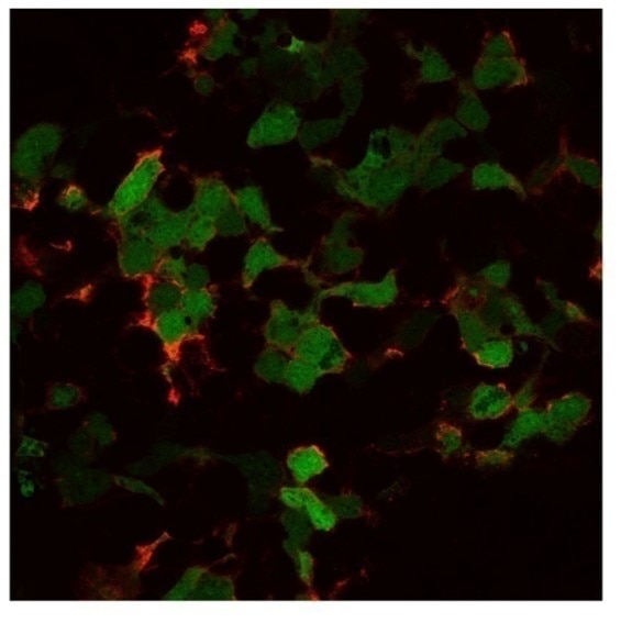
![Immunocytochemistry/Immunofluorescence: GFP Antibody (9F9.F9) [NB600-597] - GFP-transducted 293T cells. Image from verified customer review.](http://images.novusbio.com/fullsize/GFP-Antibody-9F9-F9-Immunocytochemistry-Immunofluorescence-NB600-597-img0019.jpg "Immunocytochemistry/Immunofluorescence: GFP Antibody (9F9.F9) [NB600-597] - GFP-transducted 293T cells. Image from verified customer review.")
| Reactivity | All-NASpecies Glossary |
| Applications | WB, DB, ELISA, Flow, Func, ICC/IF, IHC, IP, WB, KD |
| Clone | 9F9.F9 |
| Clonality | Monoclonal |
| Host | Mouse |
| Conjugate | Unconjugated |
| Description | GFP Monoclonal Antibody was prepared from tissue culture supernatant by Protein A affinity chromatography. Assay by Immunoelectrophoresis resulted in a single precipitin arc against anti-Mouse Serum Store this antibody at -20C prior to opening. Aliquot contents and freeze at -20C or below for extended storage. Avoid cycles of freezing and thawing. Centrifuge product if not completely clear after standing at room temperature. This product is stable for several weeks at 4C as an undiluted liquid. Dilute only prior to immediate use. |
| Immunogen | Recombinant Green Fluorescent Protein (GFP) fusion protein corresponding to the full length amino acid sequence (246 aa) derived from the jellyfish Aequorea victoria. |
| Specificity | Reactivity is observed against recombinant Green Fluorescent Protein from Aequorea victoria by both Western blot and ELISA. No reaction is seen against RFP. |
| Isotype | IgG1 Kappa |
| Clonality | Monoclonal |
| Host | Mouse |
| Purity | Protein A purified |
| Innovator's Reward | Test in a species/application not listed above to receive a full credit towards a future purchase. |
| Dilutions |
|
|
| Application Notes | This product is designed to detect enhanced GFP and GFP containing recombinant proteins. Tested in ELISA, IP, and WB and suitable in FACS, IHC, IF. This antibody can be used to detect GFP by ELISA (sandwich or capture) for the direct binding of antigen. Biotin conjugated monoclonal anti-GFP is well suited to titrate GFP in a sandwich ELISA in combination with polyclonal anti-GFP as the capture antibody. Only use the monoclonal form for the detection of enhanced or recombinant GFP. Polyclonal anti-GFP detects all variants of GFP tested to date. The biotin conjugated detection antibody is typically used with streptavidin conjugated HRP or other streptavidin conjugates. The use of polyclonal anti-GFP results in significant amplification of signal when fluorochrome conjugated polyclonal anti-GFP is used relative to the fluorescence of GFP alone. For immunoblotting use either alkaline phosphatase or peroxidase conjugated anti-GFP to detect GFP or GFP containing proteins on western blots. Optimal titers for applications should be determined by the researcher. Use in In vitro assay reported in scientific literature (PMID 23125142). Use in dot blot reported in scientific literature (PMID 24497578). Use in immunoprecipitation reported in scientific literature (PMID 28198010). Knockdown validation (Wolf et al). Use in immunoblotting reported in scientific literature (PMID:31727786). |
|
| Reviewed Applications |
|
|
| Publications |
|
| Storage | Store at -20C. Avoid freeze-thaw cycles. |
| Buffer | 0.02 M Potassium Phosphate, 0.15 M Sodium Chloride, pH 7.2 |
| Preservative | 0.01% Sodium Azide |
| Purity | Protein A purified |
| Images | Ratings | Applications | Species | Date | Details | ||||||||||
|---|---|---|---|---|---|---|---|---|---|---|---|---|---|---|---|
Enlarge |
reviewed by:
Feng Li |
WB | Human | 11/18/2019 |
Summary
|
||||||||||
Enlarge |
reviewed by:
Verified Customer |
ICC | Human | 05/10/2018 |
Summary
Comments
|
||||||||||

Enlarge |
reviewed by:
Verified Customer |
WB | Other | 01/26/2015 |
Summary
|
Secondary Antibodies |
Isotype Controls |
Research Areas for GFP Antibody (NB600-597)Find related products by research area.
|
|
Successful Transplantation of Friedreich Ataxia Induced Pluripotent Stem Cell (iPSC)-Derived Sensory Neurons in Dorsal Root Ganglia of Adult Rodents Jamshed Arslan, Pharm D, PhD The dorsal root ganglia (DRG) are a collection of cell bodies of sensory nerves carrying sensory information – including nociception, mechanoreception and proprioception – from periphera... Read full blog post. |
|
Autophagy and RAS signaling: Clinical implications By Christina Towers, PhD The cellular recycling process known as autophagy is currently being targeted in over 60 clinical trials focused on treating different types of cancer1. To date, the only autophagy-targeted ... Read full blog post. |
|
Neurovascular signaling for repair enhances brain metastasis By Jamshed Arslan, Pharm. D., PhD. Stroke is a leading cause of mortality and morbidity worldwide. Cellular players – neurons, astrocytes, endothelial and stromal cells – involved in post-stroke repair t... Read full blog post. |
|
Read full blog post. |
|
How to visualize autophagy by microscopy By Christina Towers, PhD Autophagy is a recycling process that relies on the formation of a unique organelle termed an autophagosome. An elegant way to monitor autophagy is through various microscopy techniques to... Read full blog post. |
|
Toll-like receptors in the intestinal epithelial cells By Jamshed Arslan, Pharm. D., PhD. Toll-like receptors (TLRs) are microbe-sensing proteins that act as first responders to danger signals. TLRs help the intestinal epithelial cells (IECs) recognize commensal bacteria ... Read full blog post. |
|
Read full blog post. |
|
Animal Models to Study Autophagy By Christina Towers, PhD What is autophagy?Autophagy is the catabolic process that degrades cytoplasmic material via the lysosome. The process of macroautophagy was originally characterized in yeast, where the... Read full blog post. |
|
The use of a GFP antibody for research applications in transgenic C. elegans, GFP tagged yeast and porcine model GFP, or green fluorescent protein, is a chemiluminescent protein derived from Aequorea jellyfish that was first discovered by Osamu Shimomura. It was soon after established that the emission spectra of GFP was right around 509nm, or the ultraviol... Read full blog post. |
|
GFP - Be Green! Green fluorescence protein (GFP) is a 27KD protein derived from the jellyfish Aquorea victoria that emits a green light (emission peak at a wavelength of 509 nm) when excited by blue light (excitation peak at a wavelength of 395 nm). GFP is a highly v... Read full blog post. |

The concentration calculator allows you to quickly calculate the volume, mass or concentration of your vial. Simply enter your mass, volume, or concentration values for your reagent and the calculator will determine the rest.
5 | |
4 | |
3 | |
2 | |
1 |
| Feng Li 11/18/2019 |
||
| Application: | WB | |
| Species: | Human |
| Verified Customer 05/10/2018 |
||
| Application: | ICC | |
| Species: | Human |
| Verified Customer 01/26/2015 |
||
| Application: | WB | |
| Species: | Other |


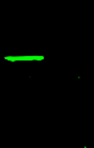
![Western Blot: GFP Antibody (9F9.F9) [NB600-597] - Lane 1: Molecular Weight Markers. Lane 2: GFP. Load: 10ng per lane. Primary antibody: Ms Anti-GFP antibody at 1:1000 for overnight at 4C. Secondary antibody: Mouse HRP secondary antibody at 1:40,000 for 30 min at RT. Block: 5% BLOTTO overnight at 4C. Predicted/Observed size: 27 kDa, 27 kDa for epitope tag GFP. Other band(s): None.](http://images.novusbio.com/fullsize/GFP-Antibody-9F9-F9-Western-Blot-NB600-597-img0017.jpg "Western Blot: GFP Antibody (9F9.F9) [NB600-597] - Lane 1: Molecular Weight Markers. Lane 2: GFP. Load: 10ng per lane. Primary antibody: Ms Anti-GFP antibody at 1:1000 for overnight at 4C. Secondary antibody: Mouse HRP secondary antibody at 1:40,000 for 30 min at RT. Block: 5% BLOTTO overnight at 4C. Predicted/Observed size: 27 kDa, 27 kDa for epitope tag GFP. Other band(s): None.")
![Western Blot: GFP Antibody (9F9.F9) [NB600-597] - All lanes have picogram amount recombinant GFP protein spiked into HeLa cell-derived lysates Lane 1: 64pg 2pg Lane 3: 16pg Lane 4: 8pg Lane 5: 4pg Lane 6: 2pg Lane 7: 1g Lane 8: 0pg. Primary antibody: anti-GFP monoclonal antibody at 1:400 for overnight at 4C. Secondary antibody: HRP-conjugated anti-Mouse IgG was performed at a dilution of 1:20,000 for 1h at 4C. Block: TTBS supplemented with 1% BSA for 1 h at 4C. Predicted/Observed size: 27 kDa for GFP. Other b&(s): none.](http://images.novusbio.com/fullsize/GFP-Antibody-9F9-F9-Western-Blot-NB600-597-img0015.jpg "Western Blot: GFP Antibody (9F9.F9) [NB600-597] - All lanes have picogram amount recombinant GFP protein spiked into HeLa cell-derived lysates Lane 1: 64pg 2pg Lane 3: 16pg Lane 4: 8pg Lane 5: 4pg Lane 6: 2pg Lane 7: 1g Lane 8: 0pg. Primary antibody: anti-GFP monoclonal antibody at 1:400 for overnight at 4C. Secondary antibody: HRP-conjugated anti-Mouse IgG was performed at a dilution of 1:20,000 for 1h at 4C. Block: TTBS supplemented with 1% BSA for 1 h at 4C. Predicted/Observed size: 27 kDa for GFP. Other b&(s): none.")
![Immunohistochemistry: GFP Antibody (9F9.F9) [NB600-597] - Used 1:5000 As referenced in: Lu S-M, Tremblay M-E` , King IL, Qi J, Reynolds HM, et al. (2011) HIV-1 Tat-Induced Microgliosis and Synaptic Damage via Interactions between Peripheral and Central Myeloid Cells. PLoS ONE 6(9): e23915. doi:10.1371/journal.pone.0023915 Image using the Biotin format of this antibody.](http://images.novusbio.com/fullsize/GFP-Antibody-9F9-F9-Immunohistochemistry-NB600-597-img0020.jpg "Immunohistochemistry: GFP Antibody (9F9.F9) [NB600-597] - Used 1:5000 As referenced in: Lu S-M, Tremblay M-E` , King IL, Qi J, Reynolds HM, et al. (2011) HIV-1 Tat-Induced Microgliosis and Synaptic Damage via Interactions between Peripheral and Central Myeloid Cells. PLoS ONE 6(9): e23915. doi:10.1371/journal.pone.0023915 Image using the Biotin format of this antibody.")
![Western Blot: GFP Antibody (9F9.F9) [NB600-597] - Expression of GFP in CHO-K1 cells trasfected with GFP. Image from verified customer review.](http://images.novusbio.com/fullsize/GFP-Antibody-9F9-F9-Western-Blot-NB600-597-img0007.jpg "Western Blot: GFP Antibody (9F9.F9) [NB600-597] - Expression of GFP in CHO-K1 cells trasfected with GFP. Image from verified customer review.")

. Lane 1: Opal Prestained Molecular Weight Marker")

 using a 1:1000 dilution of HRP-conjugated Anti-Mouse IgG Secondary Antibody (Catalog # HAF007). This experiment was conducted under reducing conditions and using Immunoblot Buffer Group 1.")
![Flow Cytometry: Mouse IgG1 Kappa Isotype Control (P3.6.2.8.1) [NBP1-43319] - Analysis of Alexa Fluor (R) 700 conjugate of NBP1-43319. Human buffy coat stained with mouse IgG1 Kappa isotype control. Flow cytometry image submitted by a verified customer review.](https://images.novusbio.com/images/Mouse-IgG1-Kappa-Light-Chain-Isotype-Control-P3-6-2-8-1-Flow-Cytometry-NBP1-43319-img0002.jpg "Flow Cytometry: Mouse IgG1 Kappa Isotype Control (P3.6.2.8.1) [NBP1-43319] - Analysis of Alexa Fluor (R) 700 conjugate of NBP1-43319. Human buffy coat stained with mouse IgG1 Kappa isotype control. Flow cytometry image submitted by a verified customer review.")


