Disease and disorder research has been conducted in relation to the Regulation Of Appetite Pathway and Obesity, Nervousness, Pituitary Diseases, Diabetes Mellitus, Hyperphagia. The study of the Regulation Of Appetite Pathway has been mentioned in research publications which can be found using our bioinformatics tool below. The Regulation Of Appetite Pathway has been researched in relation to Secretion, Energy Homeostasis, Pathogenesis, Feeding Behavior, Glucose Homeostasis. The Regulation Of Appetite Pathway complements our catalog of research reagents including antibodies and ELISA kits against GHRELIN, GROWTH HORMONE, GLUCAGON-LIKE PEPTIDE-1, CCK, CNR1.
Top Research Reagents
We have 2464 products for the study of the Regulation Of Appetite Pathway that can be applied to Flow Cytometry, Immunocytochemistry/ Immunofluorescence, Immunohistochemistry, Western Blot from our catalog of antibodies and ELISA kits.


![Western Blot: RIPK2 Antibody (6F7) [H00008767-M02] - RIPK2 monoclonal antibody (M02), clone 6F7. Analysis of RIPK2 expression in NIH/3T3.](https://images.novusbio.com/fullsize/RIPK2-Antibody-6F7-Western-Blot-H00008767-M02-img0013.jpg)
![Immunohistochemistry-Paraffin: RIPK2 Antibody (6F7) [H00008767-M02] - Analysis of monoclonal antibody to RIPK2 on formalin-fixed paraffin-embedded human prostate. Antibody concentration 1.2 ug/ml.](https://images.novusbio.com/fullsize/RIPK2-Antibody-6F7-Immunohistochemistry-Paraffin-H00008767-M02-img0008.jpg)
![Immunocytochemistry/Immunofluorescence: Cannabinoid R1/CB1/CNR1 Antibody [NLS32] - Analysis of anti-CNR1 / CB1 antibody with HEK293 human embryonic kidney cells.](https://images.novusbio.com/fullsize/Cannabinoid-R1-CB1-CNR1-Antibody-Immunocytochemistry-Immunofluorescence-NLS32-img0007.jpg)
![Immunohistochemistry-Paraffin: Cannabinoid R1/CB1/CNR1 Antibody [NLS32] - Brain, substantia nigra](https://images.novusbio.com/fullsize/Cannabinoid-R1-CB1-CNR1-Antibody-Immunohistochemistry-Paraffin-NLS32-img0013.jpg)
![Immunohistochemistry: Neuropeptide Y Antibody [NBP1-46535] - Low magnification images showing the double staining pattern of NHE1 with neuropeptide NPY. Scale bar: 100um. Insets: High magnification images showing individual cells with double staining. Scale bar: 10um. Asterisks mark Hoechst-stained nuclei. Image collected and cropped by CiteAb from the following publication (nature.com/articles/s41598-019-42872-w), licensed under a CC-BY license.](https://images.novusbio.com/fullsize/Neuropeptide-Y-Antibody-Immunohistochemistry-NBP1-46535-img0001.jpg)
![Immunocytochemistry/ Immunofluorescence: Neuropeptide Y Antibody [NBP1-46535] - NHE1 distribution (A) & colocalisation with markers for specific cell types (B–D) & major inputs (E, F, G). (A) NHE1 immunoreactivity is distributed throughout the rostrocaudal axis of the SCN (encircled by the dotted lines). Scale bar: 200 µm. OC: optic chiasm. 3 V: third ventricle. (B–G1) Low magnification images showing the double staining pattern of NHE1 with neuropeptides NP2 (B), GRP (C), & VIP (D) as well as markers for afferent inputs vGluT2 (E), NPY (F), & SERT (G1). Scale bar: 100 µm. Insets: High magnification images showing individual cells with double staining. Scale bar: 10 µm. Asterisks mark Hoechst-stained nuclei. (G2) High magnification image showing high degree of colocalisation (yellow) between NHE1 (green) & SERT (red). Scale bar: 10 µm. Image collected & cropped by CiteAb from the following publication (https://pubmed.ncbi.nlm.nih.gov/31015514), licensed under a CC-BY license. Not internally tested by Novus Biologicals.](https://images.novusbio.com/fullsize/nbp1-46535_goat-polyclonal-neuropeptide-y-antibody-310202416161848.jpg)
![Immunohistochemistry-Paraffin: Peptide YY Antibody [NBP1-80865] - Analysis in human colon and liver tissues using NBP1-80865 antibody. Corresponding Peptide YY RNA-seq data are presented for the same tissues.](https://images.novusbio.com/fullsize/Peptide-YY-Antibody-Immunohistochemistry-Paraffin-NBP1-80865-img0007.jpg)
![Immunohistochemistry-Paraffin: Peptide YY Antibody [NBP1-80865] - Staining of human liver shows low expression as expected.](https://images.novusbio.com/fullsize/Peptide-YY-Antibody-Immunohistochemistry-Paraffin-NBP1-80865-img0004.jpg)
![Western Blot: gamma-glutamyl hydrolase Antibody [NBP1-90927] - Analysis in control (vector only transfected HEK293T lysate) and GGH over-expression lysate (Co-expressed with a C-terminal myc-DDK tag (3.1 kDa) in mammalian HEK293T cells).](https://images.novusbio.com/fullsize/gamma-glutamyl-hydrolase-Antibody-Western-Blot-NBP1-90927-img0006.jpg)
![Immunohistochemistry-Paraffin: gamma-glutamyl hydrolase Antibody [NBP1-90927] - Staining of human skeletal muscle shows very weak cytoplsmic positivity in myocytes.](https://images.novusbio.com/fullsize/gamma-glutamyl-hydrolase-Antibody-Immunohistochemistry-Paraffin-NBP1-90927-img0010.jpg)
![Western Blot: Ghrelin Receptor/GHSR Antibody [NBP2-16655] - Various whole cell extracts (30 ug) were separated by 10% SDS-PAGE, and the membrane was blotted with GHS-R1 antibody [N1], N-term diluted at 1:500. The HRP-conjugated anti-rabbit IgG antibody (NBP2-19301) was used to detect the primary antibody.](https://images.novusbio.com/fullsize/Ghrelin-Receptor-GHSR-Antibody-Western-Blot-NBP2-16655-img0009.jpg)
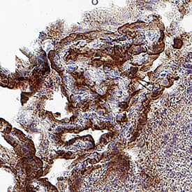
![Immunohistochemistry-Paraffin: Ghrelin Receptor/GHSR Antibody [NBP2-16655] - CL1-0 xenograft, using GHS-R1 antibody at 1:100 dilution. Antigen Retrieval: Citrate buffer, pH 6.0, 15 min.](https://images.novusbio.com/fullsize/GHSR-Antibody-Immunohistochemistry-Paraffin-NBP2-16655-img0002.jpg)
![Immunohistochemistry: POMC Antibody [NB100-1533] - Representative confocal images of POMC in POMC-transfected WT and Sel1L-/- N2a cells. White arrows point to POMC-containing secretory granules, while yellow arrows point to perinuclear POMC. KDEL marks the ER. Representative data from at least 2 independent experiments are shown. Image collected and cropped by CiteAb from the following publication (jci.org/articles/view/96420), licensed under a CC-BY license.](https://images.novusbio.com/fullsize/POMC-Antibody-Immunohistochemistry-NB100-1533-img0008.jpg)
![Flow Cytometry: POMC Antibody [NB100-1533] - Flow cytometric analysis of paraformaldehyde fixed A431 cells (blue line), permeabilized with 0.5% Triton. Primary incubation 1hr (10 ug/mL) followed by Alexa Fluor 488 secondary antibody (1 ug/mL). IgG control: Unimmunized goat IgG (black line) followed by Alexa Fluor 488 secondary antibody.](https://images.novusbio.com/fullsize/POMC-Antibody-Flow-Cytometry-NB100-1533-img0006.jpg)


![N/A Adiponectin/Acrp30 [HRP]](https://images.novusbio.com/fullsize2/DATA_Adiponectin_DRP300_ELISA_812.jpg)
![N/A Adiponectin/Acrp30 [HRP]](https://images.novusbio.com/fullsize2/DATA_Adiponectin_DRP300_ELISA_813.jpg)

![Western Blot: Corticotropin Releasing Factor Antibody (2B11) [H00001392-M02] - Analysis of CRH expression in transfected 293T cell line by CRH monoclonal antibody (M02), clone 2B11.Lane 1: CRH transfected lysate(21.4 KDa).Lane 2: Non-transfected lysate.](https://images.novusbio.com/fullsize/Corticotropin-Releasing-Factor-Antibody-2B11-Western-Blot-H00001392-M02-img0002.jpg)
![Sandwich ELISA: Corticotropin Releasing Factor Antibody (2B11) [H00001392-M02] - Detection limit for recombinant GST tagged CRH is 0.3 ng/ml as a capture antibody.](https://images.novusbio.com/fullsize/Corticotropin-Releasing-Factor-Antibody-2B11-Sandwich-ELISA-H00001392-M02-img0001.jpg)
![N/A Leptin/OB [HRP]](https://images.novusbio.com/fullsize2/DATA_Leptin_MOB00_ELISA_1010.jpg)
![N/A Leptin/OB [HRP]](https://images.novusbio.com/fullsize2/Leptin_MOB00_ELISA_2738.jpg)
