| Submit your blog on Nucleobase Transport to be featured! |
| Submit your event on Nucleobase Transport to be featured! |
![Western Blot: NRAS Antibody (6S6F3) [NBP3-16473] - Analysis of extracts of Rat lung, using NRAS Rabbit mAb (NBP3-16473) at 1:1000 dilution. Secondary antibody: HRP Goat Anti-Rabbit IgG (H+L) at 1:10000 dilution. Lysates/proteins: 25ug per lane. Blocking buffer: 3% nonfat dry milk in TBST. Detection: ECL Basic Kit. Exposure time: 1s.](https://images.novusbio.com/fullsize/NRAS-Antibody-6S6F3-Western-Blot-NBP3-16473-img0002.jpg)
![Immunocytochemistry/Immunofluorescence: NRAS Antibody (6S6F3) [NBP3-16473] - Immunofluorescence analysis of U-2 OS cells using NRAS Rabbit mAb (NBP3-16473) at dilution of 1:100 (40x lens). Blue: DAPI for nuclear staining.](https://images.novusbio.com/fullsize/NRAS-Antibody-6S6F3-Immunocytochemistry-Immunofluorescence-NBP3-16473-img0001.jpg)
Rabbit Monoclonal
Species Human, Mouse, Rat
Applications WB, ICC/IF
|
|
![Western Blot: Xanthine Oxidase Antibody (0M8E7) [NBP3-16735] - Western blot analysis of extracts of various cell lines, using Xanthine Oxidase (XDH) Rabbit mAb (NBP3-16735) at 1:1000 dilution. Secondary antibody: HRP Goat Anti-Rabbit IgG (H+L) at 1:10000 dilution. Lysates/proteins: 25ug per lane. Blocking buffer: 3% nonfat dry milk in TBST. Detection: ECL Basic Kit. Exposure time: 60s.](https://images.novusbio.com/fullsize/Xanthine-Oxidase-Antibody-0M8E7-Western-Blot-NBP3-16735-img0005.jpg)
![Immunocytochemistry/ Immunofluorescence: Xanthine Oxidase Antibody (0M8E7) [NBP3-16735] - Confocal imaging of paraffin-embedded Human liver using Xanthine Oxidase Rabbit mAb followed by a further incubation with Cy3 Goat Anti-Rabbit IgG (H+L) . DAPI was used for nuclear staining (Blue). Objective: 40x. Perform high pressure antigen retrieval with 0.01 M citrate buffer (pH 6.0) prior to IF staining.](https://images.novusbio.com/fullsize/nbp3-16735_rabbit-xanthine-oxidase-mab-0m8e7-52202511411449.jpg)
Rabbit Monoclonal
Species Human, Mouse, Rat
Applications WB, ELISA, ICC/IF
|
|
Mouse Monoclonal
Applications IHC
|
|
![SDS-Page: Recombinant Human SLC23A2 Protein [H00009962-P01] - 12.5% SDS-PAGE Stained with Coomassie Blue.](https://images.novusbio.com/fullsize/qc_test-H00009962-P01-1.jpg)
Species Human
Applications WB, ELISA, PA
|
|
![Western Blot: PSMA7 Antibody (1A10-3G12) [H00005688-M01] - Analysis of PSMA7 expression in transfected 293T cell line by PSMA7 monoclonal antibody (M01), clone 1A10-3G12.Lane 1: PSMA7 transfected lysate (Predicted MW: 27.9 KDa).Lane 2: Non-transfected lysate.](https://images.novusbio.com/fullsize/PSMA7-Antibody-1A10-3G12-Western-Blot-H00005688-M01-img0018.jpg)
![Immunocytochemistry/Immunofluorescence: PSMA7 Antibody (1A10-3G12) [H00005688-M01] - Analysis of monoclonal antibody to PSMA7 on HeLa cell. Antibody concentration 1 ~ 10 ug/ml.](https://images.novusbio.com/fullsize/PSMA7-Antibody-1A10-3G12-Immunocytochemistry-Immunofluorescence-H00005688-M01-img0009.jpg)
Mouse Monoclonal
Species Human
Applications WB, ELISA, ICC/IF
| 1 Review 2 Publications |
|
![Immunohistochemistry: PD-ECGF/Thymidine Phosphorylase Antibody (PGF 44C) [NB100-2737] - Immunohistochemical staining of thymidine phosphorylase (TP) expression and related markers in benign lymph node. In benign lymph node, TP expression highlights the meshwork of macrophages (arrow in inset; 400x) and endothelial cells (arrowhead in inset; 400x). Images are from an Olympus BX41 microscope (Olympus Corp., Tokyo, Japan) processed with Qcapture Pro 5.1 (QImaging, Surrey, BC, Canada). Image collected and cropped by CiteAb from the following publication (https://www.hindawi.com/journals/ah/2011/875135/) licensed under a CC-BY license.](https://images.novusbio.com/fullsize/PD-ECGF-Thymidine-Phosphorylase-Antibody-PGF-44C-Immunohistochemistry-NB100-2737-img0003.jpg)
![Immunohistochemistry: PD-ECGF/Thymidine Phosphorylase Antibody (PGF 44C) [NB100-2737] - Immunohistochemical staining of thymidine phosphorylase (TP) expression and related markers in benign lymph node in lymph node with malignant mycosis fungoides/Sezary syndrome cells (LN-MF). CD21 is negative in this case of LN-MF. Images are from an Olympus BX41 microscope (Olympus Corp., Tokyo, Japan) processed with Qcapture Pro 5.1 (QImaging, Surrey, BC, Canada). Image collected and cropped by CiteAb from the following publication (https://www.hindawi.com/journals/ah/2011/875135/) licensed under a CC-BY license.](https://images.novusbio.com/fullsize/PD-ECGF-Thymidine-Phosphorylase-Antibody-PGF-44C-Immunohistochemistry-NB100-2737-img0008.jpg)
Mouse Monoclonal
Species Human
Applications WB, ICC/IF, IHC
| 1 Publication |
|
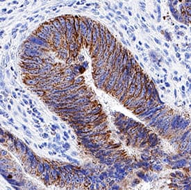
![Immunohistochemistry: ENT2 Antibody (7B2J3) [NBP3-33206] - Immunohistochemistry analysis of paraffin-embedded Rat ovary using ENT2 Rabbit mAb at dilution of 1:100 (40x lens). Microwave antigen retrieval performed with 0.01M Tris/EDTA Buffer (pH 9.0) prior to IHC staining.](https://images.novusbio.com/fullsize/nbp3-33206_rabbit-ent2-mab-7b2j3-91220241519542.jpg)
![Immunohistochemistry: ENT2 Antibody (7B2J3) [NBP3-33206] - Immunohistochemistry analysis of paraffin-embedded Human esophageal cancer using ENT2 Rabbit mAb at dilution of 1:100 (40x lens). Microwave antigen retrieval performed with 0.01M Tris/EDTA Buffer (pH 9.0) prior to IHC staining.](https://images.novusbio.com/fullsize/nbp3-33206_rabbit-ent2-mab-7b2j3-91220241519554.jpg)
Rabbit Monoclonal
Species Human, Mouse, Rat
Applications ELISA, ICC/IF, IHC
|
|
![Western Blot: HPRT Antibody [NBP1-33527] - Various whole cell extracts (30 ug) were separated by 12% SDS-PAGE, and the membrane was blotted with HPRT antibody diluted at 1:500. HRP-conjugated anti-rabbit IgG antibody was used to detect the primary antibody.](https://images.novusbio.com/fullsize/HPRT-Antibody-Western-Blot-NBP1-33527-img0015.jpg)
![Immunocytochemistry/Immunofluorescence: HPRT Antibody [NBP1-33527] - Paraformaldehyde-fixed HeLa, using antibody at 1:200 dilution.](https://images.novusbio.com/fullsize/HPRT-Antibody-Immunocytochemistry-Immunofluorescence-NBP1-33527-img0002.jpg)
Rabbit Polyclonal
Species Human, Mouse, Rat
Applications WB, Simple Western, ICC/IF
| 4 Publications |
|
![Immunohistochemistry-Paraffin: ENT1 Antibody [NBP1-84838] - Staining of human colon shows strong cytoplasmic positivity in glandular cells.](https://images.novusbio.com/fullsize/ENT1-Antibody-Immunohistochemistry-Paraffin-NBP1-84838-img0004.jpg)
Rabbit Polyclonal
Species Human
Applications IHC, IHC-P
| 1 Publication |
|
![Western Blot: Complement C6 Antibody [NBP1-91803] - Lane 1: Marker [kDa] 250, 130, 95, 72, 55, 36, 28, 17, 10. Lane 2: Human cell line RT-4. Lane 3: Human cell line U-251MG sp. Lane 4: Human plasma (IgG/HSA depleted)](https://images.novusbio.com/fullsize/Complement-C6-Antibody-Western-Blot-NBP1-91803-img0004.jpg)
![Immunohistochemistry-Paraffin: Complement C6 Antibody [NBP1-91803] - Staining of human prostate shows strong granular cytoplasmic and membranous positivity in glandular cells.](https://images.novusbio.com/fullsize/Complement-C6-Antibody-Immunohistochemistry-Paraffin-NBP1-91803-img0002.jpg)
Rabbit Polyclonal
Species Human
Applications WB, IHC, IHC-P
|
|
![Immunohistochemistry-Paraffin: SLC23A1 Antibody [NBP2-13318] - Analysis in human small intestine and pancreas tissues. Corresponding SLC23A1 RNA-seq data are presented for the same tissues.](https://images.novusbio.com/fullsize/SLC23A1-Antibody-Immunohistochemistry-Paraffin-NBP2-13318-img0006.jpg)
![Immunohistochemistry-Paraffin: SLC23A1 Antibody [NBP2-13318] - Staining of human pancreas shows no positivity as expected.](https://images.novusbio.com/fullsize/SLC23A1-Antibody-Immunohistochemistry-Paraffin-NBP2-13318-img0008.jpg)
Rabbit Polyclonal
Species Human
Applications WB, IHC, IHC-P
| 3 Publications |
|
![Western Blot: RENT1/UPF1/hUPF1 Antibody [NB100-368] - Samples: Whole cell lysate (50 ug) from HeLa and mouse NIH3T3 cells prepared using NETN lysis buffer. Antibody: Affinity purified goat anti-RENT1 antibody used for WB at 0.4 ug/ml. Detection: Chemiluminescence with an exposure time of 30 seconds.](https://images.novusbio.com/fullsize/RENT1-UPF1-hUPF1-Antibody-Western-Blot-NB100-368-img0004.jpg)
![Western Blot: RENT1/UPF1/hUPF1 Antibody [NB100-368] - Whole cell lysate (30 ug) from lymphoblastoid cell lines (EBV-immotalized B-cell from a normal individual). Antibody used at 1 ug/ml.](https://images.novusbio.com/fullsize/RENT1-UPF1-hUPF1-Antibody-Western-Blot-NB100-368-img0003.jpg)
Goat Polyclonal
Species Human, Mouse
Applications WB
| 10 Publications |
|


Sheep Polyclonal
Species Human
Applications WB, ICC
| 2 Publications |
|

Sheep Polyclonal
Species Human, Mouse
Applications WB
|
|
![Immunohistochemistry-Paraffin: LAMC2 Antibody (CL2980) [NBP2-42388] - Staining in human fallopian tube and liver tissues. Corresponding LAMC2 RNA-seq data are presented for the same tissues.](https://images.novusbio.com/fullsize/LAMC2-Antibody-CL2980-Immunohistochemistry-Paraffin-NBP2-42388-img0023.jpg)
![Western Blot: LAMC2 Antibody (CL2980) [NBP2-42388] - Analysis in A-431 cells transfected with control siRNA, target specific siRNA probe #1 and #2, using Anti-LAMC2 antibody. Remaining relative intensity is presented. Loading control: Anti-GAPDH.](https://images.novusbio.com/fullsize/LAMC2-Antibody-CL2980-Western-Blot-NBP2-42388-img0017.jpg)
Mouse Monoclonal
Species Human
Applications WB, ICC/IF, IHC
| 4 Publications |
|
![Western Blot: HRAS Antibody [NBP2-42864] - Various whole cell extracts (30 ug) were separated by 12% SDS-PAGE, and the membrane was blotted with H-RAS antibody diluted at 1:1000. The HRP-conjugated anti-rabbit IgG antibody (NBP2-19301) was used to detect the primary antibody.](https://images.novusbio.com/fullsize/HRAS-Antibody-Western-Blot-NBP2-42864-img0011.jpg)
![Immunocytochemistry/Immunofluorescence: HRAS Antibody [NBP2-42864] - Analysis of paraformaldehyde-fixed HeLa, using H-Ras antibody at 1:200 dilution.](https://images.novusbio.com/fullsize/HRAS-Antibody-Immunocytochemistry-Immunofluorescence-NBP2-42864-img0001.jpg)
Rabbit Polyclonal
Species Human, Mouse, Rat
Applications WB, ICC/IF, IHC
| 2 Publications |
|
![Immunohistochemistry-Paraffin: UPF2 Antibody [NBP2-94094] - Paraffin-embedded rat kidney using UPF2 .](https://images.novusbio.com/fullsize/UPF2-Antibody-Immunohistochemistry-Paraffin-NBP2-94094-img0001.jpg)
![Immunoprecipitation: UPF2 Antibody - BSA Free [UPF2] - Immunoprecipitation analysis of 300ug extracts of 293T cells using 3ug UPF2 Rabbit pAb . Western blot was performed from the immunoprecipitate using UPF2 Rabbit pAb at a dilition of 1:1000.](https://images.novusbio.com/fullsize/nbp2-94094_rabbit-polyclonal-upf2-antibody-52202516321424.jpg)
Rabbit Polyclonal
Species Human, Rat
Applications WB, IHC, IHC-P
|
|
![Dual RNAscope ISH-IHC: TLR4 Antibody (76B357.1) [NB100-56566] - FFPE tissue sections of human tonsil were probed for TLR4 mRNA (ACD RNAScope Probe, ACD catalog # 311281; Fast Red chromogen, ACD catalog # 322750). Adjacent tissue section was processed for immunohistochemistry using Mouse Monoclonal (Novus Biologicals catalog # NB100-56566) at 5ug/mL with 1 hour incubation at room temperature followed by incubation with anti-mouse IgG VisUCyte HRP Polymer Antibody (Catalog # VC001) and DAB chromogen (yellow-brown). Tissue was counterstained with hematoxylin (blue). Specific staining was localized to lymphocytes.](https://images.novusbio.com/fullsize/TLR4-Antibody-76B357-1-Dual-RNAscope-ISH-IHC-NB100-56566-img0036.jpg)
![Flow Cytometry: TLR4 Antibody (76B357.1) [NB100-56566] - Analysis using the Alexa Fluor (R) 647 conjugate of NBP2-27149. TLR4 expression on monocytes from human peripheral blood. PBMC were stained in a 2 color flow test, with CD14 PE version of this antibody and 1 ug of either isotype control (left) or TLR4-Alexa Fluor 647 (right). PPI negative, CD14+ cells were gated for analysis.](https://images.novusbio.com/fullsize/TLR4-Antibody-76B357-1-Flow-Cytometry-NB100-56566-img0024.jpg)
Mouse Monoclonal
Species Human, Mouse, Rat
Applications WB, Simple Western, DB
| 5 Reviews 145 Publications |
|
