| Submit your blog on Xeroderma Pigmentosum, Complementation Group C to be featured! |
| Submit your event on Xeroderma Pigmentosum, Complementation Group C to be featured! |
![Western Blot: Dihydrofolate Reductase/DHFR Antibody (2B10) [H00001719-M01] - Folic acid treatment restores endothelial specific DHFR expression and activity in Ang II-infused apoE null mice.DHFR expression (A, n?=?8-11) and activity (B, n?=?5-7) measured from endothelial cells (ECs) and EC-denuded aortas isolated from apoE null mice after 4 weeks of infusion. Data show that DHFR expression and activity was restored in FA treated animals specifically in the endothelial cells. eNOS was stained to show that isolation/removal of ECs from the vessels was successful. *p<0.05. Image collected and cropped by CiteAb from the following publication (//dx.plos.org/10.1371/journal.pone.0088899) licensed under a CC-BY license.](https://images.novusbio.com/fullsize/Dihydrofolate-Reductase-DHFR-Antibody-2B10-Western-Blot-H00001719-M01-img0017.jpg)
![Immunocytochemistry/Immunofluorescence: Dihydrofolate Reductase/DHFR Antibody (2B10) [H00001719-M01] - Analysis of monoclonal antibody to DHFR on HeLa cell. Antibody concentration 10 ug/ml.](https://images.novusbio.com/fullsize/Dihydrofolate-Reductase-DHFR-Antibody-2B10-Immunocytochemistry-Immunofluorescence-H00001719-M01-img0012.jpg)
Mouse Monoclonal
Species Human, Mouse, Rat
Applications WB, ELISA, ICC/IF
| 12 Publications |
|
Mouse Monoclonal
Applications IHC
|
|
![Western Blot: iNOS Antibody [NB300-605] - Analysis of iNOS was performed by loading 20 ug of RAW264 whole cell lysate untreated (left lane) or stimulated with LPS at 1 ug/mL for 16 hours (right lane) and 10 uL of PageRuler Plus Prestained Protein Ladder onto a 4-20% Tris-Glycine polyacrylamide gel. Proteins were transferred to a nitrocellulose membrane and blocked with 5% Milk in TBST for at least 1 hour. The membrane was probed with an iNOS Rabbit polyclonal antibody at a dilution of 1:1000 overnight at 4C on a rocking platform, washed in TBST, and probed with a Goat anti-Rabbit IgG (H+L) Secondary Antibody, HRP conjugate at a dilution of 1:1000 for 1 hour. Chemiluminescent detection was performed using SuperSignal West Pico.](https://images.novusbio.com/fullsize/iNOS-Antibody-Western-Blot-NB300-605-img0015.jpg)
![Immunohistochemistry-Paraffin: iNOS Antibody [NB300-605] - Immunohistochemistry was performed on normal deparaffinized human Lung tissue.](https://images.novusbio.com/fullsize/iNOS-Antibody-Immunohistochemistry-Paraffin-NB300-605-img0010.jpg)
Rabbit Polyclonal
Species Human, Mouse, Rat
Applications WB, Flow, IB
| 3 Reviews 141 Publications |
|
![Western Blot: XPG Antibody [NB100-74611] - Detection of human ERCC5/XPG by western blot. Samples: Whole cell lysate (50 ug) from Jurkat, HeLa, and HEK293T cells prepared using NETN lysis buffer. Antibody: Affinity purified rabbit anti-ERCC5/XPG antibody NB100-74611 used for WB at 0.04 ug/ml. Detection: Chemiluminescence with an exposure time of 3 minutes.](https://images.novusbio.com/fullsize/XPG-Antibody-Western-Blot-NB100-74611-img0010.jpg)
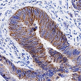
![Immunohistochemistry-Paraffin: XPG Antibody [NB100-74611] - FFPE section of human colon carcinoma. Antibody: Affinity purified rabbit anti-ERCC5/XPG used at a dilution of 1:200 (1ug/ml). Detection: DAB](https://images.novusbio.com/fullsize/XPG-Antibody-Immunohistochemistry-Paraffin-NB100-74611-img0011.jpg)
Rabbit Polyclonal
Species Human
Applications WB, ICC/IF, IHC
| 5 Publications |
|
![Western Blot: GTF2H1 Antibody (1F12-1B5) [H00002965-M01] - Analysis of GTF2H1 expression in transfected 293T cell line by GTF2H1 monoclonal antibody (M01), clone 1F12-1B5.Lane 1: GTF2H1 transfected lysate(62 KDa).Lane 2: Non-transfected lysate.](https://images.novusbio.com/fullsize/GTF2H1-Antibody-1F12-1B5-Western-Blot-H00002965-M01-img0016.jpg)
![Immunocytochemistry/Immunofluorescence: GTF2H1 Antibody (1F12-1B5) [H00002965-M01] - Analysis of monoclonal antibody to GTF2H1 on HeLa cell. Antibody concentration 10 ug/ml.](https://images.novusbio.com/fullsize/GTF2H1-Antibody-1F12-1B5-Immunocytochemistry-Immunofluorescence-H00002965-M01-img0009.jpg)
Mouse Monoclonal
Species Human
Applications WB, ELISA, ICC/IF
| 2 Publications |
|
![Western Blot: GRP75/HSPA9B/Mortalin Antibody (9F8) [NBP1-47801] - Analysis of extracts from 9 different cell lines: (HepG2: human; HeLa: human; SVT2: mouse; A549: human; COS7: monkey; Jurkat: human; MDCK: canine; PC12: rat; MCF7: human).](https://images.novusbio.com/fullsize/GRP75-HSPA9B-Mortalin-Antibody-9F8-Western-Blot-NBP1-47801-img0022.jpg)
![Immunocytochemistry/Immunofluorescence: GRP75/HSPA9B/Mortalin Antibody (9F8) [NBP1-47801] - Staining of COS7 cells transiently transfected by pCMV6-ENTRY GRP75/HSPA9B/Mortalin.](https://images.novusbio.com/fullsize/GRP75-HSPA9B-Mortalin-Antibody-9F8-Immunocytochemistry-Immunofluorescence-NBP1-47801-img0014.jpg)
Mouse Monoclonal
Species Human, Mouse, Rat
Applications WB, Flow, ICC/IF
| 1 Publication |
|
![Western Blot: p53 Antibody (PAb 240) [NB200-103] - Analysis of p53 in MCF7 and HeLa lystates. Image courtesy of anonymous customer product review.](https://images.novusbio.com/fullsize/p53-Antibody-PAb-240-Western-Blot-NB200-103-img0001.jpg)
![Immunocytochemistry/Immunofluorescence: p53 Antibody (PAb 240) [NB200-103] - PC12 cells were fixed in 4% paraformaldehyde for 10 minutes and permeabilized in 0.5% Triton X-100 in PBS for 5 minutes. The cells were incubated with anti-p53 Antibody (PAb 240) NB200-103 at 2 ug/ml overnight at 4C and detected with an anti-mouse Dylight 488 (Green) at a 1:1000 dilution for 60 minutes. Nuclei were counterstained with DAPI (Blue). Cells were imaged using a 40X objective.](https://images.novusbio.com/fullsize/p53-Antibody-PAb-240-Immunocytochemistry-Immunofluorescence-NB200-103-img0013.jpg)
Mouse Monoclonal
Species Human, Mouse, Rat
Applications WB, ELISA, Flow
| 3 Reviews 43 Publications |
|
Mouse Monoclonal
Species Human, Mouse, Rat
Applications WB, ELISA, Flow
| 3 Reviews 43 Publications |
|
![Immunohistochemistry-Paraffin: hHR23b Antibody [NBP1-89699] - Staining of human colon, kidney, liver and testis using Anti-RAD23B antibody NBP1-89699 (A) shows similar protein distribution across tissues to independent antibody NBP1-89698 (B).](https://images.novusbio.com/fullsize/hHR23b-Antibody-Immunohistochemistry-Paraffin-NBP1-89699-img0011.jpg)
![Western Blot: hHR23b Antibody [NBP1-89699] - Analysis in human cell line RH-30.](https://images.novusbio.com/fullsize/hHR23b-Antibody-Western-Blot-NBP1-89699-img0010.jpg)
Rabbit Polyclonal
Species Human
Applications WB, ICC/IF, IHC
| 1 Publication |
|
Rabbit Polyclonal
Species Human
Applications WB, ICC/IF, IHC
| 1 Publication |
|
![Western Blot: S5a/Angiocidin Antibody [NBP2-19952] - Immunoblots of parental HCT116 cells and two clonal cell lines (clone 14 and clone 13) generated by CRISPR-mediated truncation of hRpn10. Image collected and cropped by CiteAb from the following publication (nature.com/articles/s41467-020-15073-7), licensed under a CC-BY license.](https://images.novusbio.com/fullsize/S5a-Angiocidin-Antibody-Western-Blot-NBP2-19952-img0004.jpg)
![Immunocytochemistry/Immunofluorescence: Proteasome 19S S5A Antibody [NBP2-19952] - Immunofluorescence analysis of paraformaldehyde-fixed A549, using antibody at 1:200 dilution.](https://images.novusbio.com/fullsize/Proteasome-19S-S5A-Antibody-Immunocytochemistry-Immunofluorescence-NBP2-19952-img0002.jpg)
Rabbit Polyclonal
Species Human
Applications WB, ICC/IF, KD
| 2 Publications |
|
![Western Blot: MDM2/HDM2 Antibody (SMP14) [NB100-2736] - Daudi whole cell protein was separated by SDS-PAGE on a 7.5% gel and transferred to PVDF membrane. The membrane was probed with anti-MDM2 antibody at 2 ug/ml and detected with an anti-mouse HRP secondary antibody using chemiluminescence.](https://images.novusbio.com/fullsize/MDM2-HDM2-Antibody-SMP14-Western-Blot-NB100-2736-img0001.jpg)
![Immunohistochemistry-Paraffin: MDM2/HDM2 Antibody (SMP14) [NB100-2736] - Analysis of tissue section of human breast cancer xenograft using MDM2/HDM2 antibody (clone SMP14) at 1:100 dilution. Several of the cancer cells developed a strong nuclear with weak cytoplasmic immunostaining of MDM2.](https://images.novusbio.com/fullsize/MDM2-HDM2-Antibody-SMP14-Immunohistochemistry-Paraffin-NB100-2736-img0003.jpg)
Mouse Monoclonal
Species Human, Mouse, Rat
Applications WB, IHC, IHC-Fr
| 1 Review 22 Publications |
|
![Immunohistochemistry-Paraffin: Glucagon R/GCGR Antibody [NLS4257] - Brain, Glioblastoma](https://images.novusbio.com/fullsize/Glucagon-R-GCGR-Antibody-Immunohistochemistry-Paraffin-NLS4257-img0005.jpg)
![Immunohistochemistry-Paraffin: Glucagon R/GCGR Antibody [NLS4257] - Human liver tissue after heat-induced antigen retrieval.](https://images.novusbio.com/fullsize/Glucagon-R-GCGR-Antibody-Immunohistochemistry-Paraffin-NLS4257-img0001.jpg)
Rabbit Polyclonal
Species Human, Bovine, Canine
Applications IHC, IHC-P
|
|
![Western Blot: E2F-4 Antibody [NBP1-21374] - Detection of Human E2F4 by Western Blot. Samples: Whole cell lysate from HeLa (5, 15, 50 ug) and 293T (50ug) cells prepared using NETN lysis buffer. Antibody: Affinity purified rabbit anti-E2F4 antibody NBP1-21374 used for WB at 0.1 ug/ml. Detection: Chemiluminescence with an exposure time of 30 seconds.](https://images.novusbio.com/fullsize/E2F-4-Antibody-Western-Blot-NBP1-21374-img0039.jpg)
![Immunohistochemistry-Paraffin: E2F-4 Antibody [NBP1-21374] - Sample: FFPE section of human prostate carcinoma. Antibody: Affinity purified rabbit anti-E2F4 used at a dilution of 1:1,000 (1ug/ml). Detection: DAB](https://images.novusbio.com/fullsize/E2F-4-Antibody-Immunohistochemistry-NBP1-21374-img0037.jpg)
Rabbit Polyclonal
Species Human
Applications WB, IHC, IHC-P
| 3 Publications |
|


Mouse Monoclonal
Species Human
Applications WB, IHC, IP
| 40 Publications |
|

Goat Polyclonal
Species Human
Applications WB
| 7 Publications |
|

Goat Polyclonal
Species Human
Applications WB
| 1 Publication |
|
![Western Blot: ERCC8 Antibody (2G1) [NBP2-45519] - Analysis of HEK293T cells were transfected with the pCMV6-ENTRY control (Left lane) or pCMV6-ENTRY ERCC8.](https://images.novusbio.com/fullsize/ERCC8-Antibody-2G1-Western-Blot-NBP2-45519-img0006.jpg)
![Immunohistochemistry: ERCC8 Antibody (2G1) [NBP2-45519] - Analysis of Human bladder tissue. (Heat-induced epitope retrieval by 1mM EDTA in 10mM Tris buffer (pH8.5) at 120C for 3 min)](https://images.novusbio.com/fullsize/ERCC8-Antibody-2G1-Immunohistochemistry-NBP2-45519-img0005.jpg)
Mouse Monoclonal
Species Human, Mouse, Rat
Applications WB, IHC, IHC-P
|
|
![Western Blot: REEP5 Antibody (4D2) [NBP2-45945] - Analysis of HEK293T cells were transfected with the pCMV6-ENTRY control (Left lane) or pCMV6-ENTRY REEP5.](https://images.novusbio.com/fullsize/REEP5-Antibody-4D2-Western-Blot-NBP2-45945-img0008.jpg)
![Immunohistochemistry: REEP5 Antibody (4D2) [NBP2-45945] - Analysis of Carcinoma of Human thyroid tissue. (Heat-induced epitope retrieval by 10mM citric buffer, pH6.0, 120C for 3min)](https://images.novusbio.com/fullsize/REEP5-Antibody-4D2-Immunohistochemistry-NBP2-45945-img0007.jpg)
Mouse monoclonal
Species Human, Mouse, Rat
Applications WB, Flow, IHC
|
|
![Western Blot: LXR beta/NR1H2 Antibody [NB100-74457] - 25 ug of rat liver (Lane 1), mouse liver (Lane 2) and Hela (Lane 3) cell lysates.](https://images.novusbio.com/fullsize/LXR-beta-NR1H2-Antibody-Western-Blot-NB100-74457-img0004.jpg)
![Immunocytochemistry/Immunofluorescence: LXR beta/NR1H2 Antibody [NB100-74457] - Analysis of LXR beta (green) showing positive staining in the nuclear envelope of C2C12 cells (right) compared with a negative control in the absence of primary antibody (left).](https://images.novusbio.com/fullsize/LXR-beta-NR1H2-Antibody-Immunocytochemistry-Immunofluorescence-NB100-74457-img0003.jpg)
Rabbit Polyclonal
Species Human, Mouse, Rat
Applications WB, ICC/IF
| 7 Publications |
|
Rabbit Polyclonal
Species Human, Mouse, Rat
Applications WB, ICC/IF
| 7 Publications |
|
![Western Blot: Rad23 Antibody (3E3F4) [NBP2-52549] - Analysis using RAD23A mAb against human RAD23A (AA: 1-363) recombinant protein. (Expected MW is 42.5 kDa)](https://images.novusbio.com/fullsize/hHR23A-Antibody-3E3F4-Western-Blot-NBP2-52549-img0004.jpg)
![Immunohistochemistry-Paraffin: Rad23 Antibody (3E3F4) [NBP2-52549] - Analysis of paraffin-embedded esophageal cancer tissues using RAD23A mouse mAb with DAB staining.](https://images.novusbio.com/fullsize/hHR23A-Antibody-3E3F4-Immunohistochemistry-Paraffin-NBP2-52549-img0003.jpg)
Mouse Monoclonal
Species Human
Applications WB, ELISA, Flow
|
|
Mouse Monoclonal
Species Human
Applications WB, ELISA, Flow
|
|
![Western Blot: XPC Antibody (3.26) [NB100-477] - DNA repair protein expression in tumors. A. Western blot determination of p53, XPC, XPA, ERCC1 and MSH2 in tumors. GAPDH is loading control. Image collected and cropped by CiteAb from the following publication (//ovarianresearch.biomedcentral.com/articles/10.1186/1757-2215-4-9) licensed under a CC-BY license.](https://images.novusbio.com/fullsize/XPC-Antibody-3-26-Western-Blot-NB100-477-img0007.jpg)
![Western Blot: XPC Antibody (3.26) [NB100-477] - HeLa whole cell lysate detected using rabbit polyclonal .](https://images.novusbio.com/fullsize/XPC-Antibody-3-26-Western-Blot-NB100-477-img0004.jpg)
Mouse Monoclonal
Species Human, Mouse
Applications WB, ICC/IF, IHC
| 1 Review 21 Publications |
|
Mouse Monoclonal
Species Human, Mouse
Applications WB, ICC/IF, IHC
| 1 Review 21 Publications |
|
![Western Blot: p14ARF/CDKN2A Antibody [NB200-111] - Inhibition of AKT decreases p53mut stability. T24 cells were transfected with non-targeting control, AKT1, or p14ARF siRNA. Cells were treated with NCS348884 (4 i1/4M), Nutlin3A (5 i1/4M) or DMSO as indicated. Whole cell lysates were probed with the indicated antibodies. Image collected and cropped by Citeab from the following publication (AKT regulates NPM dependent ARF localization and p53mut stability in tumors. <i>Oncotarget</i> (2014)) licensed under a CC-BY license.](https://images.novusbio.com/fullsize/p14ARF-CDKN2A-Antibody-Western-Blot-NB200-111-img0012.jpg)
![Immunohistochemistry: p14ARF/CDKN2A Antibody [NB200-111] - Inhibition of AKT modulates p53 stability in-vivo and synergizes with ionizing radiation to inhibit tumor growth( Sections of PSN1 xenografts treated with three consecutive doses of MK-2206 (60 mg/kg). Sections of PSN1 xenografts and in-vitro PSN1 cells fixed and stained with anti-NPM (red) and anti-p14ARF (green). Image collected and cropped by Citeab from the following publication (AKT regulates NPM dependent ARF localization and p53mut stability in tumors. Oncotarget (2014)) licensed under a CC-BY license.](https://images.novusbio.com/fullsize/p14ARF-CDKN2A-Antibody-Immunocytochemistry-Immunofluorescence-NB200-111-img0011.jpg)
Rabbit Polyclonal
Species Human, Mouse
Applications WB, Flow, ICC/IF
| 2 Reviews 18 Publications |
|
![Western Blot: DDB1 Antibody (JU32-35) [NBP2-75465] - Analysis of DDB1 on PC-12 cell lysates with Rabbit anti-DDB1 antibody at 1/500 dilution. Lysates/proteins at 10 ug/Lane. Predicted band size: 127 kDa Observed band size: 127 kDa Exposure time: 2 minutes; 8% SDS-PAGE gel. Proteins were transferred to a PVDF membrane and blocked with 5% NFDM/TBST for 1 hour at room temperature. The primary antibody at 1/500 dilution was used in 5% NFDM/TBST at room temperature for 2 hours. Goat Anti-Rabbit IgG - HRP Secondary Antibody at 1:200,000 dilution was used for 1 hour at room temperature.](https://images.novusbio.com/fullsize/DDB1-Antibody-JU32-35-Western-Blot-NBP2-75465-img0009.jpg)
![Immunocytochemistry/Immunofluorescence: DDB1 Antibody (JU32-35) [NBP2-75465] - Staining DDB1 in SH-SY5Y cells (green). The nuclear counter stain is DAPI (blue). Cells were fixed in paraformaldehyde, permeabilised with 0.25% Triton X100/PBS.](https://images.novusbio.com/fullsize/DDB1-Antibody-JU32-35-Immunocytochemistry-Immunofluorescence-NBP2-75465-img0003.jpg)
Rabbit Monoclonal
Species Human, Mouse, Rat
Applications WB, ICC/IF, IHC
| 3 Publications |
|
![Immunocytochemistry/Immunofluorescence: nNOS Antibody [NB100-858] - Action potentials were evoked with depolarizing current pulses. (A) Neuron from colonic specimen of non-treated patient fired 1 action potential in response to a depolarizing current. (A') Intracellular injection of carboxyfluorescein during recording confirmed Dogiel type I, uniaxonal morphology. (A](https://images.novusbio.com/fullsize/nNOS-Antibody-Immunocytochemistry-Immunofluorescence-NB100-858-img0005.jpg)
![Immunohistochemistry-Paraffin: nNOS Antibody [NB100-858] - Staining of paraffin embedded Human Cortex. Steamed antigen retrieval with citrate buffer pH 6, AP-staining. Antibody at 2.5 ug/mL.](https://images.novusbio.com/fullsize/nNOS-Antibody-Immunohistochemistry-Paraffin-NB100-858-img0006.jpg)
Goat Polyclonal
Species Human, Mouse, Rat
Applications WB, Flow, ICC/IF
| 1 Review 15 Publications |
|
![Western Blot: Centrin 2 Antibody [NBP2-92653] - Analysis of extracts of various cell lines, using CETN2 antibody at 1:1000 dilution.Secondary antibody: HRP Goat Anti-Rabbit IgG (H+L) at 1:10000 dilution.Lysates/proteins: 25ug per lane. Blocking buffer: 3% nonfat dry milk in TBST.Detection: ECL Basic Kit. Exposure time: 180s.](https://images.novusbio.com/fullsize/Centrin-2-Antibody-Western-Blot-NBP2-92653-img0003.jpg)
![Western Blot: Centrin 2 Antibody [NBP2-92653] - Analysis of extracts from normal (control) and CETN2 knockout (KO) HeLa cells, using CETN2 antibody at 1:1000 dilution. Secondary antibody: HRP Goat Anti-Rabbit IgG (H+L) at 1:10000 dilution. Lysates/proteins: 25ug per lane.Blocking buffer: 3% nonfat dry milk in TBST. Detection: ECL Basic Kit. Exposure time: 180s.](https://images.novusbio.com/fullsize/Centrin-2-Antibody-Knockout-Validated-NBP2-92653-img0005.jpg)
Rabbit Polyclonal
Species Human, Mouse, Rat
Applications WB, ICC/IF, KO
|
|
![Western Blot: DP1 Antibody [NBP2-92787] - Analysis of extracts of various cell lines, using DP1 at 1:3000 dilution. Secondary antibody: HRP Goat Anti-Rabbit IgG (H+L) at 1:10000 dilution. Lysates/proteins: 25ug per lane. Blocking buffer: 3% nonfat dry milk in TBST. Detection: ECL Basic Kit . Exposure time: 90s.](https://images.novusbio.com/fullsize/DP1-Antibody-Western-Blot-NBP2-92787-img0003.jpg)
![Immunohistochemistry-Paraffin: DP1 Antibody [NBP2-92787] - Paraffin-embedded human lung using DP1 .](https://images.novusbio.com/fullsize/DP1-Antibody-Immunohistochemistry-Paraffin-NBP2-92787-img0002.jpg)
Rabbit Polyclonal
Species Human, Mouse
Applications WB, IHC, IHC-P
|
|
![Western Blot: XPB Antibody [NB100-61060] - Whole cell lysate (15 ug) from HeLa, 293T, Jurkat, mouse TCMK-1, and mouse NIH3T3 cells prepared using NETN lysis buffer. Antibody: Affinity purified rabbit antiERCC3 antibody used for WB at 0.1 ug/mL. Detection: Chemiluminescence with an exposure time of 3 minutes.](https://images.novusbio.com/fullsize/XPB-Antibody-Western-Blot-NB100-61060-img0006.jpg)
![Immunoprecipitation: XPB Antibody [NB100-61060] - Detection of human ERCC3 by western blot of immunoprecipitates. Samples: Whole cell lysate (1.0 mg per IP reaction; 20% of IP loaded) from HEK293T cells prepared using NETN lysis buffer. Antibodies: Affinity purified rabbit anti-ERCC3 antibody NB100-61060 used for IP at 3 ug per reaction. ERCC3 was also immunoprecipitated by rabbit anti-ERCC3 antibody NB100-61059. For blotting immunoprecipitated ERCC3, NB100-61060 was used at 1 ug/ml. Detection: Chemiluminescence with an exposure time of 3 minutes.](https://images.novusbio.com/fullsize/XPB-Antibody-Immunoprecipitation-NB100-61060-img0005.jpg)
Rabbit Polyclonal
Species Human, Mouse, Rabbit
Applications WB, IP, ChIP
| 1 Review 3 Publications |
|
