| Submit your blog on Nephrocalcinosis to be featured! |
| Submit your event on Nephrocalcinosis to be featured! |

Species Human
Applications WB, ELISA, PA
|
|
![Western Blot: Serpin A8/Angiotensinogen Antibody [NBP1-30027] - Analysis of Angiotensinogen in human kidney lysate.](https://images.novusbio.com/fullsize/Serpin-A8-Angiotensinogen-Antibody-Western-Blot-NBP1-30027-img0013.jpg)
![Immunocytochemistry/Immunofluorescence: Serpin A8/Angiotensinogen Antibody [NBP1-30027] - HepG2 cells were fixed for 10 minutes using 10% formalin and then permeabilized for 5 minutes using 1X TBS + 0.5% Triton-X100. The cells were incubated with anti-Angiotensinogen at 10 ug/ml overnight at 4C and detected with an anti-rabbit Dylight 488 (Green) at a 1:500 dilution. Alpha tubulin (DM1A) NB100-690 was used as a co-stain at a 1:1000 dilution and detected with an anti-mouse Dylight 550 (Red) at a 1:500 dilution. Nuclei were counterstained with DAPI (Blue). Cells were imaged using a 40X objective.](https://images.novusbio.com/fullsize/Serpin-A8-Angiotensinogen-Antibody-Immunocytochemistry-Immunofluorescence-NBP1-30027-img0014.jpg)
Rabbit Polyclonal
Species Human, Mouse, Rat
Applications WB, ICC/IF, IHC
| 11 Publications |
|
![Western Blot: SLC12A3 Antibody [NBP1-44270] - Western blot analysis of Rat tissue lysates showing detection of SLC12A3 protein using Rabbit Anti-SLC12A3 Polyclonal Antibody (NBP1-44270). Primary Antibody: Rabbit Anti-SLC12A3 Polyclonal Antibody (NBP1-44270) at 1:1000.](https://images.novusbio.com/fullsize/SLC12A3-Antibody-Western-Blot-NBP1-44270-img0009.jpg)
![Immunohistochemistry: SLC12A3 Antibody [NBP1-44270] - Immunohistochemistry analysis using Rabbit Anti-SLC12A3 Polyclonal Antibody (NBP1-44270). Tissue: kidney tissue. Species: Rat. Primary Antibody: Rabbit Anti-SLC12A3 Polyclonal Antibody (NBP1-44270) at 1:200. Secondary Antibody: FITC Goat Anti-Rabbit (green).](https://images.novusbio.com/fullsize/SLC12A3-Antibody-Immunohistochemistry-NBP1-44270-img0010.jpg)
Rabbit Polyclonal
Species Human, Mouse, Rat
Applications WB, EM, ICC/IF
| 1 Publication |
|
![Western Blot: CLCN5 Antibody [NBP1-69123] - Rat Brain Antibody Dilution: 1.0 ug/ml](https://images.novusbio.com/fullsize/CLCN5-Antibody-Western-Blot-NBP1-69123-img0009.jpg)
![Western Blot: CLCN5 Antibody [NBP1-69123] - Mouse Brain lysate, concentration 0.2-1 ug/ml.](https://images.novusbio.com/fullsize/CLCN5-Antibody-Western-Blot-NBP1-69123-img0002.jpg)
Rabbit Polyclonal
Species Mouse, Rat
Applications WB
|
|
![Immunohistochemistry-Paraffin: NKCC2/SLC12A1 Antibody [NBP1-80993] - Analysis in human kidney and pancreas tissues using NBP1-80993 antibody. Corresponding SLC12A1 RNA-seq data are presented for the same tissues.](https://images.novusbio.com/fullsize/NKCC2-SLC12A1-Antibody-Immunohistochemistry-Paraffin-NBP1-80993-img0018.jpg)
![Immunohistochemistry-Paraffin: NKCC2/SLC12A1 Antibody [NBP1-80993] - Staining of human cerebellum, kidney, liver and pancreas using Anti-SLC12A1 antibody NBP1-80993 (A) shows similar protein distribution across tissues to independent antibody NBP1-82559 (B).](https://images.novusbio.com/fullsize/NKCC2-SLC12A1-Antibody-Immunohistochemistry-Paraffin-NBP1-80993-img0013.jpg)
Rabbit Polyclonal
Species Human
Applications WB, ICC/IF, IHC
| 1 Publication |
|
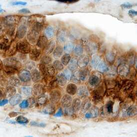
![Immunohistochemistry-Paraffin: KCNJ1 Antibody [NBP1-82874] -Analysis in human kidney and pancreas tissues using NBP1-82874 antibody. Corresponding KCNJ1 RNA-seq data are presented for the same tissues.](https://images.novusbio.com/fullsize/antibody/nbp1-82874_rabbit-polyclonal-kcnj1-antibody-immunohistochemistry-paraffin-14620248590..jpg)
![Immunohistochemistry-Paraffin: KCNJ1 Antibody [NBP1-82874] - Staining of human kidney shows high expression.](https://images.novusbio.com/fullsize/KCNJ1-Antibody-Immunohistochemistry-Paraffin-NBP1-82874-img0003.jpg)
Rabbit Polyclonal
Species Human, Mouse, Rat
Applications ICC/IF, IHC, IHC-P
| 4 Publications |
|
![Immunohistochemistry-Paraffin: AGXT Antibody [NBP1-89200] - Analysis in human liver and pancreas tissues. Corresponding AGXT RNA-seq data are presented for the same tissues.](https://images.novusbio.com/fullsize/AGXT-Antibody-Immunohistochemistry-Paraffin-NBP1-89200-img0018.jpg)
![Immunohistochemistry-Paraffin: AGXT Antibody [NBP1-89200] - Staining of human endometrium, fallopian tube, skin and testis using Anti-NEK1 antibody (A) NBP1-89200 shows similar protein distribution across tissues to independent antibody NBP2-34198 (B).](https://images.novusbio.com/fullsize/AGXT-Antibody-Immunohistochemistry-Paraffin-NBP1-89200-img0023.jpg)
Rabbit Polyclonal
Species Human, Mouse
Applications WB, Simple Western, IHC
| 3 Publications |
|

Species Human
Applications WB, ELISA, PA
|
|


Goat Polyclonal
Species Mouse
Applications WB, IHC, IP
| 13 Publications |
|


Sheep Polyclonal
Species Mouse
Applications WB, IHC
| 8 Publications |
|


Goat Polyclonal
Species Mouse
Applications WB, IHC, ELISA(Cap)
| 2 Reviews 203 Publications |
|
Mouse Monoclonal
Species Human
Applications IHC
| 3 Publications |
|


Mouse Monoclonal
Species Human
Applications WB, Simple Western, IHC
| 3 Reviews 54 Publications |
|


Mouse Monoclonal
Species Human
Applications WB, IHC
|
|
Species Human
Applications EnzAct
|
|
![Immunohistochemistry-Paraffin: Band 3 Antibody [NBP2-49409] - Staining of human cerebral cortex shows low expression as expected.](https://images.novusbio.com/fullsize/Band-3-Antibody-Immunohistochemistry-Paraffin-NBP2-49409-img0003.jpg)
![Immunohistochemistry: Band 3 Antibody [NBP2-49409] - Staining of human spleen shows strong positivity in erythrocytes.](https://images.novusbio.com/fullsize/Band-3-Antibody-Immunohistochemistry-NBP2-49409-img0001.jpg)
Rabbit Polyclonal
Species Human
Applications IHC, IHC-P
|
|
![Western Blot: RAPGEF5 Antibody [NBP2-57362] - Analysis in human cell line RT-4, human cell line U-251 MG and human plasma.](https://images.novusbio.com/fullsize/RAPGEF5-Antibody-Western-Blot-NBP2-57362-img0002.jpg)
![Immunocytochemistry/Immunofluorescence: RAPGEF5 Antibody [NBP2-57362] - Staining of human cell line PC-3 shows localization to nucleoplasm & nuclear bodies.](https://images.novusbio.com/fullsize/RAPGEF5-Antibody-Immunocytochemistry-Immunofluorescence-NBP2-57362-img0001.jpg)
Rabbit Polyclonal
Species Human
Applications WB, ICC/IF
|
|
Mouse Monoclonal
Species Human, Rat
Applications WB, ICC/IF, IHC
| 1 Review 11 Publications |
|
![Western Blot: Calcium-sensing R/CaSR Antibody (5C10, ADD) [NB120-19347] - Total protein from human Hek293 and rat Pancreas was separated on a 7.5% gel by SDS-PAGE, transferred to PVDF membrane and blocked in 5% non-fat milk in TBST. The membrane was probed with 2.0 ug/ml anti-CaSR in block buffer and detected with an anti-mouse HRP secondary antibody using West Pico PLUS chemiluminescence detection reagent.](https://images.novusbio.com/fullsize/Calcium-sensing-R-CaSR-Antibody-5C10-ADD-Western-Blot-NB120-19347-img0014.jpg)
![Immunocytochemistry/Immunofluorescence: Calcium-sensing R/CaSR Antibody (5C10, ADD) [NB120-19347] - Analysis of primary astrocytes using Calcium-sensing R/CaSR antibody. Primary astrocytes co-stained with cilia (Arl13b, red, top left), CASR (green, bottom left), DAPI (blue, top right) and merged (bottom right). Image from verified customer review.](https://images.novusbio.com/fullsize/Calcium-sensing-R-CaSR-Antibody-5C10-ADD-Immunocytochemistry-Immunofluorescence-NB120-19347-img0015.jpg)
Mouse Monoclonal
Species Human, Mouse, Rat
Applications WB, ELISA, ICC/IF
| 1 Review 21 Publications |
|
