| Submit your blog on Intraductal Hyperplasia to be featured! |
| Submit your event on Intraductal Hyperplasia to be featured! |
![Western Blot: UVRAG Antibody [NBP1-18885] - Beclin 1 is acetylated at lysines 430 and 437. TSA and NAM increase the binding of Beclin 1 to Rubicon. Immunoprecipitation of indicated Beclin 1-binding partners with ectopically expressed Flag-Beclin 1 in HEK293T cells treated with TSA and NAM. Image collected and cropped by CiteAb from the following publication (https://www.nature.com/articles/ncomms8215), licensed under a CC-BY license.](https://images.novusbio.com/fullsize/UVRAG-Antibody-Western-Blot-NBP1-18885-img0005.jpg)
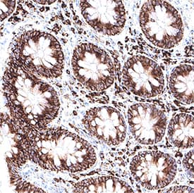
![Immunohistochemistry-Paraffin: UVRAG Antibody [NBP1-18885] - Section of human colon carcinoma. Antibody: Affinity purified rabbit anti- UVRAG used at a dilution of 1:200 (1ug/ml). Detection: DAB](https://images.novusbio.com/fullsize/UVRAG-Antibody-Immunohistochemistry-Paraffin-NBP1-18885-img0004.jpg)
Rabbit Polyclonal
Species Human
Applications WB, ICC/IF, IHC
| 4 Publications |
|
Mouse Monoclonal
Species Human
Applications WB, Simple Western, IHC
|
|
![Western Blot: ER alpha/NR3A1 Antibody (33) [NB300-560] - Analysis of recombinant ER alpha.](https://images.novusbio.com/fullsize/ER-alpha-NR3A1-Antibody-33-Western-Blot-NB300-560-img0004.jpg)
![Immunohistochemistry-Paraffin: ER alpha/NR3A1 Antibody (33) [NB300-560] - Human breast tissue (B) and magnified section (C) compared with an isotype control (A).](https://images.novusbio.com/fullsize/ER-alpha-NR3A1-Antibody-33-Immunohistochemistry-Paraffin-NB300-560-img0010.jpg)
Mouse Monoclonal
Species Human, Mouse, Rat
Applications WB, IHC, IHC-Fr
| 1 Review 11 Publications |
|
![Immunocytochemistry/Immunofluorescence: MUC-1 Antibody (SM3) [NB120-22711] - MCF7 cells were fixed for 10 minutes using 10% formalin and then permeabilized for 5 minutes using 1X PBS + 0.05% Triton-X100. The cells were incubated with anti-MUC1 [SM3] at 5 ug/ml overnight at 4C and detected with an anti-mouse IgG Dylight 488 (Green) at a 1:500 dilution. Nuclei were counterstained with DAPI (Blue). Cells were imaged using a 40X objective.](https://images.novusbio.com/fullsize/MUC-1-Antibody-SM3-Immunocytochemistry-Immunofluorescence-NB120-22711-img0004.jpg)
![Immunohistochemistry-Paraffin: MUC-1 Antibody (SM3) [NB120-22711] - IHC analysis of formalin fixed paraffin embedded tissue section of human breast cancer xenograft using MUC-1 antibody clone SM3 at 1:10 dilution. The xenograft section depicted a very specific and intense signal in the periphery of the cancer cells. The necrotic cells also developed a strong immune-positivity while the tumor stroma as well as the nuclei of cells were negative for immunostaining.](https://images.novusbio.com/fullsize/MUC-1-Antibody-SM3-Immunohistochemistry-Paraffin-NB120-22711-img0003.jpg)
Mouse Monoclonal
Species Human, Mouse
Applications ELISA, Flow, ICC/IF
| 14 Publications |
|
![Western Blot: p53 Antibody (PAb 240) [NB200-103] - Analysis of p53 in MCF7 and HeLa lystates. Image courtesy of anonymous customer product review.](https://images.novusbio.com/fullsize/p53-Antibody-PAb-240-Western-Blot-NB200-103-img0001.jpg)
![Immunocytochemistry/Immunofluorescence: p53 Antibody (PAb 240) [NB200-103] - PC12 cells were fixed in 4% paraformaldehyde for 10 minutes and permeabilized in 0.5% Triton X-100 in PBS for 5 minutes. The cells were incubated with anti-p53 Antibody (PAb 240) NB200-103 at 2 ug/ml overnight at 4C and detected with an anti-mouse Dylight 488 (Green) at a 1:1000 dilution for 60 minutes. Nuclei were counterstained with DAPI (Blue). Cells were imaged using a 40X objective.](https://images.novusbio.com/fullsize/p53-Antibody-PAb-240-Immunocytochemistry-Immunofluorescence-NB200-103-img0013.jpg)
Mouse Monoclonal
Species Human, Mouse, Rat
Applications WB, ELISA, Flow
| 3 Reviews 43 Publications |
|
![Western Blot: BRCA1 Antibody (RAY) [NB100-598] - Analysis of BRCA1 on MCF7 lysate.](https://images.novusbio.com/fullsize/BRCA1-Antibody-RAY-Western-Blot-NB100-598-img0006.jpg)
![Immunocytochemistry/Immunofluorescence: BRCA1 Antibody (RAY) [NB100-598] - BRCA (RAY) antibody was tested in MCF-7 cells with FITC (green). Nuclei and alpha-tubulin were counterstained with DAPI (blue) and DyLight 550 (red).](https://images.novusbio.com/fullsize/BRCA1-Antibody-RAY-Immunocytochemistry-Immunofluorescence-NB100-598-img0005.jpg)
Mouse Monoclonal
Species Human
Applications WB, Flow, ICC/IF
| 6 Publications |
|
![Western Blot: Androgen R/NR3C4 [p Ser213, p Ser210] Antibody (156C135.2) [NB100-56603] - Analysis using Azide Free version of NB100-56603. LNCaP cells (passage number 38) were serum-starved for 2 days. After serum starvation, cells were (A) left untreated, (B) treated with 100 ng/ml IGF-1 for 4h, or (C) incubated with 20 um LY294002 for 30 mi](https://images.novusbio.com/fullsize/Androgen-R-NR3C4-p-Ser213-p-Ser210-Antibody-156C135-2-Western-Blot-NB100-56603-img0004.jpg)
Mouse Monoclonal
Species Human, Mouse, Rat
Applications WB, IHC, IHC-P
| 14 Publications |
|
![Western Blot: Bcl-2 Antibody [NB100-56098] - Analysis of Bcl-2 in whole cell lysate from Daoy cells. Cells were transfected with (1) scrambled control siRNA or (2) Bcl-2 siRNA. Image from verified customer review.](https://images.novusbio.com/fullsize/Bcl-2-Antibody-Western-Blot-NB100-56098-img0004.jpg)
![Western Blot: Bcl-2 Antibody [NB100-56098] - EA reduced MPP+-induced dopaminergic neuronal apoptosis by increasing BDNF (brain-derived neurotrophic factor) expression and further Akt phosphorylation in the rat substantia nigra. Eight days after MPP+ administration, our Western blot results (MAB7566) show that MPP+ treatment reduced tyrosine hydroxylase and Bcl-2 expression in the ipsilateral side of the rat substantia nigra (SN), but not in the contralateral side. EA stimulation (50 Hz) enhanced mature BDNF, tyrosine hydroxylase, and Bcl-2 expression in the MPP+-treated ipsilateral side. Image collected and cropped by CiteAb from the following publication (https://www.mdpi.com/1422-0067/18/9/1846), licensed under a CC-BY license.](https://images.novusbio.com/fullsize/Bcl-2-Antibody-Western-Blot-NB100-56098-img0012.jpg)
Rabbit Polyclonal
Species Human, Mouse, Rat
Applications WB, IHC, IHC-P
| 2 Reviews 28 Publications |
|
![Western Blot: Lamin A + C Antibody (4C4) [NBP2-25151] - Analysis of different cell lysates using mouse mAb to lamin A/C, NBP2-25151, dilution 1:1,000 in green: [1] protein standard (red), [2] HeLa, [3] HEK293 [4] C6, and [5] NIH-3T3 cell lysates. Two strong bands at 74 and 65kDa correspond to the lamin A and lamin C proteins respectively, detected only in the cells of human origin. NBP2-25151 antibody failed to recognize rat or mouse proteins.](https://images.novusbio.com/fullsize/Lamin-A-+-C-Antibody-4C4-Western-Blot-NBP2-25151-img0006.jpg)
![Immunocytochemistry/Immunofluorescence: Lamin A + C Antibody (4C4) [NBP2-25151] - Analysis of HeLa cells stained with mouse mAb to lamin A/C, NBP2-25151, dilution 1:2,000 in red, and costained with rabbit pAb to HSP60, dilution 1:5,000, in green. The blue is Hoechst staining of nuclear DNA. NBP2-25151 antibody specifically labels the nuclear lamina, while the HSP60 antibody reveals protein expressed in mitochondria.](https://images.novusbio.com/fullsize/Lamin-A-+-C-Antibody-4C4-Immunocytochemistry-Immunofluorescence-NBP2-25151-img0005.jpg)
Mouse Monoclonal
Species Human, Mouse, Rat
Applications WB, ICC/IF
| 2 Publications |
|

Goat Polyclonal
Species Human
Applications WB, IHC
|
|


Goat Polyclonal
Species Human, Mouse
Applications WB, Simple Western, Flow
| 143 Publications |
|


Sheep Polyclonal
Species Human
Applications WB, Simple Western, IHC
| 2 Publications |
|


Goat Polyclonal
Species Human
Applications WB, IHC, ICC
| 1 Review 24 Publications |
|


Species Human
Applications BA
| 58 Publications |
|
![Immunocytochemistry/Immunofluorescence: TMEM37 Antibody [NBP2-47602] - Immunofluorescent staining of human cell line Hep G2 shows localization to nucleus, nucleoli fibrillar center & cytosol.](https://images.novusbio.com/fullsize/TMEM37-Antibody-Immunocytochemistry-Immunofluorescence-NBP2-47602-img0003.jpg)
![Immunohistochemistry-Paraffin: TMEM37 Antibody [NBP2-47602] - Staining in human kidney and pancreas tissues using anti-TMEM37 antibody. Corresponding TMEM37 RNA-seq data are presented for the same tissues.](https://images.novusbio.com/fullsize/TMEM37-Antibody-Immunohistochemistry-Paraffin-NBP2-47602-img0006.jpg)
Rabbit Polyclonal
Species Human
Applications ICC/IF, IHC, IHC-P
|
|
![Western Blot: Cyclin D1 Antibody (SPM587) [NBP2-32840] - HELLS expression and protein levels are modulated with YAP1/TEAD inhibition downstream of SHH signaling. Western blot showing protein levels of HELLS, cleaved caspase 3 (CC3), and Cyclin D1 in verteporfin treated CGNPs. Blot is representative of three replicates. Full-length blots are presented in Supplementary Fig. S4. Image collected and cropped by CiteAb from the following publication (https://www.nature.com/articles/s41598-019-50088-1), licensed under a CC-BY license.](https://images.novusbio.com/fullsize/Cyclin-D1-Antibody-SPM587-Western-Blot-NBP2-32840-img0006.jpg)
![Immunohistochemistry-Paraffin: Cyclin D1 Antibody (SPM587) [NBP2-32840] - Formalin-paraffin human Mantle Cell Lymphoma stained with Cyclin D1 Ab (Clone SPM587).](https://images.novusbio.com/fullsize/Cyclin-D1-Antibody-SPM587-Immunohistochemistry-Paraffin-NBP2-32840-img0004.jpg)
Mouse Monoclonal
Species Human, Mouse, Rat
Applications WB, Flow, ICC/IF
| 1 Review 9 Publications |
|
![SDS-Page: Recombinant Human PRH2 Protein [H00005555-P01] - 12.5% SDS-PAGE Stained with Coomassie Blue.](https://images.novusbio.com/fullsize/qc_test-H00005555-P01-1.jpg)
Species Human
Applications WB, ELISA, PA
|
|
