Research of Dextrocardia has been linked to Situs Inversus, Congenital Heart Defects, Transposition Of Great Vessels, Heart Septal Defects, Heart Diseases. The study of Dextrocardia has been mentioned in research publications which can be found using our bioinformatics tool below. Researched pathways related to Dextrocardia include Transposition, Pathogenesis, Localization, Transport, Cardiac Conduction. These pathways complement our catalog of research reagents for the study of Dextrocardia including antibodies and ELISA kits against SITUS INVERSUS VISCERUM, INVS, DNAH5, DNAI1, PCBD1.
Top Research Reagents
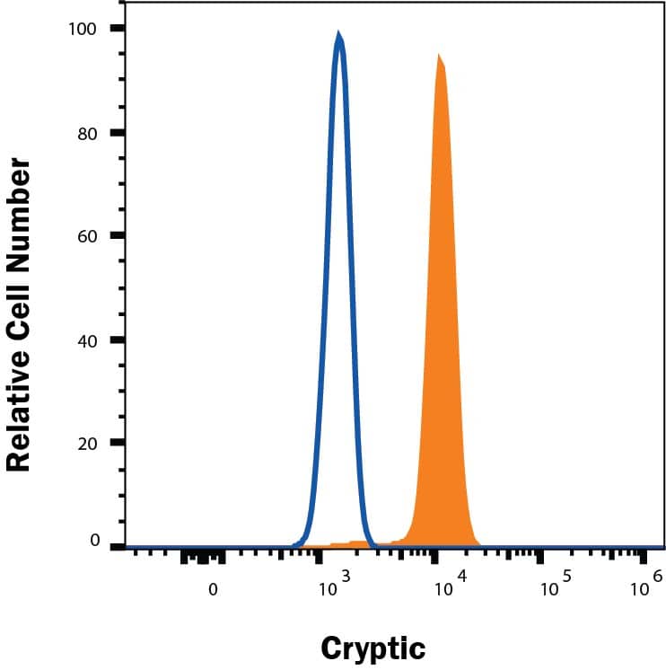
We have 432 products for the study of Dextrocardia that can be applied to Flow Cytometry, Immunocytochemistry/ Immunofluorescence, Immunohistochemistry, Western Blot from our catalog of antibodies and ELISA kits.
![Western Blot: EPB42 Antibody (2G12) [H00002038-M01] - Analysis of EPB42 expression in transfected 293T cell line by EPB42 monoclonal antibody (M01), clone 2G12.Lane 1: EPB42 transfected lysate(69.5 KDa).Lane 2: Non-transfected lysate.](https://images.novusbio.com/fullsize/EPB42-Antibody-2G12-Western-Blot-H00002038-M01-img0005.jpg)
![Immunocytochemistry/Immunofluorescence: EPB42 Antibody (2G12) [H00002038-M01] - Analysis of monoclonal antibody to EPB42 on HeLa cell . Antibody concentration 10 ug/ml.](https://images.novusbio.com/fullsize/EPB42-Antibody-2G12-Immunocytochemistry-Immunofluorescence-H00002038-M01-img0003.jpg)
![Western Blot: UVRAG Antibody [NBP1-18885] - Beclin 1 is acetylated at lysines 430 and 437. TSA and NAM increase the binding of Beclin 1 to Rubicon. Immunoprecipitation of indicated Beclin 1-binding partners with ectopically expressed Flag-Beclin 1 in HEK293T cells treated with TSA and NAM. Image collected and cropped by CiteAb from the following publication (https://www.nature.com/articles/ncomms8215), licensed under a CC-BY license.](https://images.novusbio.com/fullsize/UVRAG-Antibody-Western-Blot-NBP1-18885-img0005.jpg)
![Immunohistochemistry-Paraffin: UVRAG Antibody [NBP1-18885] - Section of human colon carcinoma. Antibody: Affinity purified rabbit anti- UVRAG used at a dilution of 1:200 (1ug/ml). Detection: DAB](https://images.novusbio.com/fullsize/UVRAG-Antibody-Immunohistochemistry-Paraffin-NBP1-18885-img0004.jpg)
![Western Blot: Pancreatic Amylase Alpha Antibody (6D4) [NBP1-47659] - Pancreatic Amylase Alpha Antibody (6D4) HEK293T cells were transfected with the pCMV6-ENTRY control (Left lane) or pCMV6-ENTRY Pancreatic Amylase(Right lane) cDNA for 48 hrs and lysed. Equivalent amounts of cell lysates (5 ug per lane) were separated by SDS-PAGE and immunoblotted with anti-Pancreatic Amylase.](https://images.novusbio.com/fullsize/Pancreatic-Amylase-Alpha-Antibody-6D4-Western-Blot-NBP1-47659-img0022.jpg)
![Immunohistochemistry-Paraffin: Pancreatic Amylase Alpha Antibody (6D4) [NBP1-47659] - Pancreatic Amylase Alpha Antibody (6D4) Staining of paraffin-embedded ovary using anti-Pancreatic Amylase mouse monoclonal antibody.](https://images.novusbio.com/fullsize/Pancreatic-Amylase-Alpha-Antibody-6D4-Immunohistochemistry-Paraffin-NBP1-47659-img0021.jpg)

![Immunohistochemistry-Paraffin: DNAH5 Antibody [NBP1-84463] - Analysis in human fallopian tube and liver tissues. Corresponding DNAH5 RNA-seq data are presented for the same tissues.](https://images.novusbio.com/fullsize/DNAH5-Antibody-Immunohistochemistry-Paraffin-NBP1-84463-img0013.jpg)
![Immunohistochemistry-Paraffin: DNAH5 Antibody [NBP1-84463] - Staining of human cerebral cortex, fallopian tube, liver and nasopharynx using Anti-DNAH5 antibody NBP1-84463 (A) shows similar protein distribution across tissues to independent antibody NBP1-84464 (B).](https://images.novusbio.com/fullsize/DNAH5-Antibody-Immunohistochemistry-Paraffin-NBP1-84463-img0016.jpg)
![Immunohistochemistry-Paraffin: DNAI1 Antibody [NBP1-84466] - Staining in human testis and prostate tissues using NBP1-84466 antibody. Corresponding DNAI1 RNA-seq data are presented for the same tissues.](https://images.novusbio.com/fullsize/DNAI1-Antibody-Immunohistochemistry-Paraffin-NBP1-84466-img0019.jpg)
![Immunohistochemistry-Paraffin: DNAI1 Antibody [NBP1-84466] - Staining of human fallopian tube, placenta, prostate and testis using Anti-DNAI1 antibody NBP1-84466 (A) shows similar protein distribution across tissues to independent antibody NBP1-84465 (B).](https://images.novusbio.com/fullsize/DNAI1-Antibody-Immunohistochemistry-Paraffin-NBP1-84466-img0015.jpg)
![Western Blot: Pallidin Antibody (1H9) [NBP2-01763] - HEK293T cells were transfected with the pCMV6-ENTRY control (Left lane) or pCMV6-ENTRY Pallidin (Right lane) cDNA for 48 hrs and lysed. Equivalent amounts of cell lysates (5 ug per lane) were separated by SDS-PAGE and immunoblotted with anti-Pallidin.](https://images.novusbio.com/fullsize/Pallidin-Antibody-1H9-Western-Blot-NBP2-01763-img0007.jpg)
![Immunohistochemistry-Paraffin: Pallidin Antibody (1H9) [NBP2-01763] - Staining of paraffin-embedded Human tonsil using anti-Pallidin mouse monoclonal antibody.](https://images.novusbio.com/fullsize/Pallidin-Antibody-1H9-Immunohistochemistry-Paraffin-NBP2-01763-img0006.jpg)
![SDS-Page: PRH1 Protein [NBP2-23368]](https://images.novusbio.com/fullsize/PRH1-Protein-SDS-Page-NBP2-23368-img0001.jpg)


![Western Blot: CS Citrate Synthase Antibody (1761) [NBP2-43648] - Non-transfected (Negative) and transfected (Positive) 293T whole cell extracts (30 ug) were separated by 10% SDS-PAGE, and the membrane was blotted with Citrate synthetase antibody diluted at 1:1000. The HRP-conjugated anti-mouse IgG antibody was used to detect the primary antibody.](https://images.novusbio.com/fullsize/CS-Citrate-Synthase-Antibody-1761-Western-Blot-NBP2-43648-img0013.jpg)
![Immunocytochemistry/Immunofluorescence: CS Citrate Synthase Antibody (1761) [NBP2-43648] - HeLa cells were fixed in 4% paraformaldehyde at RT for 15 min. Green: Citrate synthetase protein stained by Citrate synthetase antibody [1761] diluted at 1:200. Red: phalloidin, a cytoskeleton marker, diluted at 1:50. Blue: Hoechst 33342 staining.](https://images.novusbio.com/fullsize/CS-Citrate-Synthase-Antibody-1761-Immunocytochemistry-Immunofluorescence-NBP2-43648-img0012.jpg)
![Western Blot: TBX1 Antibody (OTI1C2) [NBP2-46076] - Analysis of HEK293T cells were transfected with the pCMV6-ENTRY control (Left lane) or pCMV6-ENTRY TBX1.](https://images.novusbio.com/fullsize/TBX1-Antibody-1C2-Western-Blot-NBP2-46076-img0002.jpg)
![Immunohistochemistry-Paraffin: TBX1 Antibody (OTI1C2) [NBP2-46076] - Analysis of Human lymph node tissue. (Heat-induced epitope retrieval by 1 mM EDTA in 10mM Tris, pH8.5, 120C for 3min)](https://images.novusbio.com/fullsize/TBX1-Antibody-1C2-Immunohistochemistry-NBP2-46076-img0001.jpg)
![Immunohistochemistry-Paraffin: Phenylalanine Hydroxylase Antibody [NBP2-48615] - Analysis in human liver and pancreas tissues. Corresponding Phenylalanine Hydroxylase RNA-seq data are presented for the same tissues.](https://images.novusbio.com/fullsize/Phenylalanine-Hydroxylase-Antibody-Immunohistochemistry-Paraffin-NBP2-48615-img0012.jpg)
![Immunohistochemistry-Paraffin: Phenylalanine Hydroxylase Antibody [NBP2-48615] - Staining of human cerebral cortex, kidney, liver and pancreas using Anti-Phenylalanine Hydroxylase antibody NBP2-48615 (A) shows similar protein distribution across tissues to independent antibody NBP1-80917 (B).](https://images.novusbio.com/fullsize/Phenylalanine-Hydroxylase-Antibody-Immunohistochemistry-Paraffin-NBP2-48615-img0014.jpg)
![Western Blot: PCBD1 Antibody [NBP2-93878] - Analysis of extracts of various cell lines, using PCBD1 at 1:1000 dilution.Secondary antibody: HRP Goat Anti-Rabbit IgG (H+L) at 1:10000 dilution.Lysates/proteins: 25ug per lane.Blocking buffer: 3% nonfat dry milk in TBST.Detection: ECL Basic Kit .Exposure time: 90s.](https://images.novusbio.com/fullsize/PCBD1-Antibody-Western-Blot-NBP2-93878-img0002.jpg)
![Immunocytochemistry/Immunofluorescence: PCBD1 Antibody [NBP2-93878] - Analysis of MCF7 cells using PCBD1 . Blue: DAPI for nuclear staining.](https://images.novusbio.com/fullsize/PCBD1-Antibody-Immunocytochemistry-Immunofluorescence-NBP2-93878-img0001.jpg)
![SDS-Page: Recombinant Human PRH2 Protein [H00005555-P01] - 12.5% SDS-PAGE Stained with Coomassie Blue.](https://images.novusbio.com/fullsize/qc_test-H00005555-P01-1.jpg)
