| Submit your blog on Depersonalization to be featured! |
| Submit your event on Depersonalization to be featured! |
Mouse Monoclonal
Applications IHC
|
|
![Western Blot: EPB42 Antibody (2G12) [H00002038-M01] - Analysis of EPB42 expression in transfected 293T cell line by EPB42 monoclonal antibody (M01), clone 2G12.Lane 1: EPB42 transfected lysate(69.5 KDa).Lane 2: Non-transfected lysate.](https://images.novusbio.com/fullsize/EPB42-Antibody-2G12-Western-Blot-H00002038-M01-img0005.jpg)
![Immunocytochemistry/Immunofluorescence: EPB42 Antibody (2G12) [H00002038-M01] - Analysis of monoclonal antibody to EPB42 on HeLa cell . Antibody concentration 10 ug/ml.](https://images.novusbio.com/fullsize/EPB42-Antibody-2G12-Immunocytochemistry-Immunofluorescence-H00002038-M01-img0003.jpg)
Mouse Monoclonal
Species Human
Applications WB, ELISA, ICC/IF
|
|
![Immunocytochemistry/ Immunofluorescence: AKR1C2 Antibody [NBP3-35145] - Immunofluorescence analysis of L929 cells using AKR1C2 Rabbit pAb at dilution of 1:100 (40x lens). Secondary antibody: Cy3-conjugated Goat anti-Rabbit IgG (H+L) at 1:500 dilution. Blue: DAPI for nuclear staining.](https://images.novusbio.com/fullsize/nbp3-35145_rabbit-akr1c2-pab-1012202411343318.jpg)
![Western Blot: AKR1C2 Antibody [NBP3-35145] - Western blot analysis of various lysates, using AKR1C2 Rabbit pAb at 1:2000 dilution.<br/>Secondary antibody: HRP-conjugated Goat anti-Rabbit IgG (H+L) at 1:10000 dilution.<br/>Lysates/proteins: 25ug per lane.<br/>Blocking buffer: 3% nonfat dry milk in TBST.<br/>Detection: ECL Basic Kit.<br/>Exposure time: 10s.](https://images.novusbio.com/fullsize/nbp3-35145_rabbit-akr1c2-pab-1012202411421018.jpg)
Rabbit Polyclonal
Species Human, Mouse, Rat
Applications WB, ELISA, ICC/IF
|
|
![Western Blot: Exosome component 10 Antibody [NBP1-32870] - Non-transfected (-) and transfected (+) 293T whole cell extracts (30 ug) were separated by 7.5% SDS-PAGE, and the membrane was blotted with EXOSC10 antibody diluted at 1:5000. The HRP-conjugated anti-rabbit IgG antibody (NBP2-19301) was used to detect the primary antibody.](https://images.novusbio.com/fullsize/Exosome-component-10-Antibody-Western-Blot-NBP1-32870-img0015.jpg)
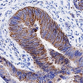
![Immunohistochemistry-Paraffin: Exosome component 10 Antibody [NBP1-32870] - Paraffin-embedded Cal27 xenograft, using antibody at 1:100 dilution.](https://images.novusbio.com/fullsize/Exosome-component-10-Antibody-Immunohistochemistry-Paraffin-NBP1-32870-img0010.jpg)
Rabbit Polyclonal
Species Human, Chicken
Applications WB, IHC, IHC-P
| 2 Reviews 3 Publications |
|
![Western Blot: Pancreatic Amylase Alpha Antibody (6D4) [NBP1-47659] - Pancreatic Amylase Alpha Antibody (6D4) HEK293T cells were transfected with the pCMV6-ENTRY control (Left lane) or pCMV6-ENTRY Pancreatic Amylase(Right lane) cDNA for 48 hrs and lysed. Equivalent amounts of cell lysates (5 ug per lane) were separated by SDS-PAGE and immunoblotted with anti-Pancreatic Amylase.](https://images.novusbio.com/fullsize/Pancreatic-Amylase-Alpha-Antibody-6D4-Western-Blot-NBP1-47659-img0022.jpg)
![Immunohistochemistry-Paraffin: Pancreatic Amylase Alpha Antibody (6D4) [NBP1-47659] - Pancreatic Amylase Alpha Antibody (6D4) Staining of paraffin-embedded ovary using anti-Pancreatic Amylase mouse monoclonal antibody.](https://images.novusbio.com/fullsize/Pancreatic-Amylase-Alpha-Antibody-6D4-Immunohistochemistry-Paraffin-NBP1-47659-img0021.jpg)
Mouse Monoclonal
Species Human
Applications WB, IHC, IHC-P
| 1 Review |
|
![Immunocytochemistry/Immunofluorescence: Aurora A Antibody [NBP1-51843] - HeLa cells were fixed and permeabilized for 10 minutes using -20C MeOH. The cells were incubated with anti- (NBP1-51843) at 2 ug/ml overnight at 4C and detected with an anti-rabbit Dylight 488 (Green) at a 1:1000 dilution for 60 minutes. Alpha tubulin (DM1A) NB100-690 was used as a co-stain at a 1:1000 dilution overnight at 4C and detected with an anti-mouse Dylight 550 (Red) at a 1:1000 dilution for 60 minutes. Nuclei were counterstained with DAPI (Blue). Cells were imaged using a 100X objective and digitally deconvolved.](https://images.novusbio.com/fullsize/Aurora-A-Antibody-Immunocytochemistry-Immunofluorescence-NBP1-51843-img0012.jpg)
![Simple Western: Aurora A Antibody [NBP1-51843] - Simple Western lane view shows a specific band for Aurora A in 0.5 mg/ml of HeLa lysate. This experiment was performed under reducing conditions using the 12-230kDa separation system.](https://images.novusbio.com/fullsize/Aurora-A-Antibody-Simple-Western-NBP1-51843-img0010.jpg)
Rabbit Polyclonal
Species Human, Mouse
Applications WB, Simple Western, ICC/IF
| 1 Review 7 Publications |
|
![Western Blot: SRPR alpha Antibody [H00006734-B02P] - Analysis of SRPR expression in human pancreas.](https://images.novusbio.com/fullsize/SRPR-alpha-Antibody-Western-Blot-H00006734-B02P-img0002.jpg)
![Immunocytochemistry/Immunofluorescence: SRPR alpha Antibody [H00006734-B02P] - Analysis of purified antibody to SRPR on HeLa cell. (antibody concentration 10 ug/ml)](https://images.novusbio.com/fullsize/SRPR-alpha-Antibody-Immunocytochemistry-Immunofluorescence-H00006734-B02P-img0001.jpg)
Mouse Polyclonal
Species Human
Applications WB, ICC/IF
| 1 Publication |
|
![Immunohistochemistry-Paraffin: p57 Kip2 Antibody [NBP1-89917] - Staining in human placenta and colon tissues using anti-CDKN1C antibody. Corresponding CDKN1C RNA-seq data are presented for the same tissues.](https://images.novusbio.com/fullsize/p57-Kip2-Antibody-Immunohistochemistry-Paraffin-NBP1-89917-img0014.jpg)
![Western Blot: p57 Kip2 Antibody [NBP1-89917] - Analysis in human placenta tissue.](https://images.novusbio.com/fullsize/p57-Kip2-Antibody-Western-Blot-NBP1-89917-img0015.jpg)
Rabbit Polyclonal
Species Human
Applications WB, ICC/IF, IHC
| 2 Publications |
|
![Western Blot: NDUFB6 Antibody [NBP1-92172] - Lane 1: Marker [kDa] 250, 130, 95, 72, 55, 36, 28, 17, 10. Lane 2: Human cell line RT-4. Lane 3: Human cell line U-251MG sp. Lane 4: Human plasma (IgG/HSA depleted). Lane 5: Human liver tissue. Lane 6: Human tonsil tissue](https://images.novusbio.com/fullsize/NDUFB6-Antibody-Western-Blot-NBP1-92172-img0006.jpg)
![Immunocytochemistry/Immunofluorescence: NDUFB6 Antibody [NBP1-92172] - Immunofluorescent staining of human cell line A-431 shows localization to nucleoplasm & mitochondria.](https://images.novusbio.com/fullsize/NDUFB6-Antibody-Immunocytochemistry-Immunofluorescence-NBP1-92172-img0007.jpg)
Rabbit Polyclonal
Species Human
Applications WB, ICC/IF, IHC
| 2 Publications |
|
![Immunocytochemistry/Immunofluorescence: NHS Antibody (6D9) [H00004810-M05] - Analysis of monoclonal antibody to NHS on HeLa cell. Antibody concentration 10 ug/ml](https://images.novusbio.com/fullsize/NHS-Antibody-6D9-Immunocytochemistry-Immunofluorescence-H00004810-M05-img0001.jpg)
![ELISA: NHS Antibody (6D9) [H00004810-M05] - Detection limit for recombinant GST tagged NHS is 0.03 ng/ml as a capture antibody.](https://images.novusbio.com/fullsize/NHS-Antibody-6D9-ELISA-H00004810-M05-img0003.jpg)
Mouse Monoclonal
Species Human
Applications WB, ELISA, ICC/IF
|
|
![Western Blot: Pallidin Antibody (1H9) [NBP2-01763] - HEK293T cells were transfected with the pCMV6-ENTRY control (Left lane) or pCMV6-ENTRY Pallidin (Right lane) cDNA for 48 hrs and lysed. Equivalent amounts of cell lysates (5 ug per lane) were separated by SDS-PAGE and immunoblotted with anti-Pallidin.](https://images.novusbio.com/fullsize/Pallidin-Antibody-1H9-Western-Blot-NBP2-01763-img0007.jpg)
![Immunohistochemistry-Paraffin: Pallidin Antibody (1H9) [NBP2-01763] - Staining of paraffin-embedded Human tonsil using anti-Pallidin mouse monoclonal antibody.](https://images.novusbio.com/fullsize/Pallidin-Antibody-1H9-Immunohistochemistry-Paraffin-NBP2-01763-img0006.jpg)
Mouse Monoclonal
Species Human, Mouse, Rat
Applications WB, Flow, IHC
|
|
![SDS-Page: PRH1 Protein [NBP2-23368]](https://images.novusbio.com/fullsize/PRH1-Protein-SDS-Page-NBP2-23368-img0001.jpg)
Species Human
Applications PAGE
|
|


Goat Polyclonal
Species Human
Applications WB, Simple Western, IHC
| 44 Publications |
|
![Western Blot: COPE Antibody [NBP2-38512] - Lane 1: Marker [kDa] 250, 130, 95, 72, 55, 36, 28, 17, 10. Lane 2: Human cell line RT-4. Lane 3: Human cell line U-251MG. Lane 4: Human Plasma. Lane 5: Human liver tissue. Lane 6: Human tonsil tissue](https://images.novusbio.com/fullsize/COPE-Antibody-Western-Blot-NBP2-38512-img0003.jpg)
![Immunocytochemistry/Immunofluorescence: COPE Antibody [NBP2-38512] - Staining of human cell line U-251 MG shows localization to the Golgi apparatus. Antibody staining is shown in green.](https://images.novusbio.com/fullsize/COPE-Antibody-Immunocytochemistry-Immunofluorescence-NBP2-38512-img0004.jpg)
Rabbit Polyclonal
Species Human, Mouse
Applications WB, ICC/IF, IHC
| 1 Publication |
|


Species Human
Applications BA
| 3 Reviews 821 Publications |
|


Species Mouse
Applications BA
| 91 Publications |
|
![Immunohistochemistry-Paraffin: Desmoplakin Antibody [NBP2-48836] - Staining in human skin and skeletal muscle tissues using anti-DSP antibody. Corresponding DSP RNA-seq data are presented for the same tissues.](https://images.novusbio.com/fullsize/Desmoplakin-Antibody-Immunohistochemistry-Paraffin-NBP2-48836-img0005.jpg)
![Immunohistochemistry-Paraffin: Desmoplakin Antibody [NBP2-48836] - Staining of human skeletal muscle shows low expression as expected.](https://images.novusbio.com/fullsize/Desmoplakin-Antibody-Immunohistochemistry-Paraffin-NBP2-48836-img0003.jpg)
Rabbit Polyclonal
Species Human, Canine
Applications ICC/IF, IHC, IHC-P
| 2 Publications |
|
![Western Blot: DPYD Antibody (7D4) [H00001806-M01] - DPYD monoclonal antibody (M01), clone 7D4. Analysis of DPYD expression in Hela S3 NE.](https://images.novusbio.com/fullsize/DPYD-Antibody-7D4-Western-Blot-H00001806-M01-img0009.jpg)
![Immunocytochemistry/Immunofluorescence: DPYD Antibody (7D4) [H00001806-M01] - Analysis of monoclonal antibody to DPYD on HeLa cell. Antibody concentration 10 ug/ml.](https://images.novusbio.com/fullsize/DPYD-Antibody-7D4-Immunocytochemistry-Immunofluorescence-H00001806-M01-img0006.jpg)
Mouse Monoclonal
Species Human
Applications WB, DB, ELISA
| 1 Publication |
|
![SDS-Page: Recombinant Human PRH2 Protein [H00005555-P01] - 12.5% SDS-PAGE Stained with Coomassie Blue.](https://images.novusbio.com/fullsize/qc_test-H00005555-P01-1.jpg)
Species Human
Applications WB, ELISA, PA
|
|
![Immunocytochemistry/Immunofluorescence: SERCA2 ATPase Antibody (2A7-A1) [NB300-581] - HeLa cells. Primary antibody at 1:100, secondary antibody: goat-anti mouse IgG AlexaFluor 568 at 1:100. ICC/IF image submitted by a verified customer review.](https://images.novusbio.com/fullsize/SERCA2-ATPase-Antibody-2A7-A1-Immunocytochemistry-Immunofluorescence-NB300-581-img0010.jpg)
![Immunohistochemistry-Paraffin: SERCA2 ATPase Antibody (2A7-A1) [NB300-581] - Both normal and cancer biopsies of deparaffinized Human skeletal muscle tissues.](https://images.novusbio.com/fullsize/SERCA2-ATPase-Antibody-2A7-A1-Immunohistochemistry-Paraffin-NB300-581-img0008.jpg)
Mouse Monoclonal
Species Human, Mouse, Rat
Applications WB, Flow, ICC/IF
| 2 Reviews 17 Publications |
|
