| Submit your blog on Deficiency Of Sulfatase to be featured! |
| Submit your event on Deficiency Of Sulfatase to be featured! |
![Western Blot: NOD2 Antibody (2D9) [NB100-524] - HCMV infection induces NOD2 mRNA and protein in HFFs and U373 cells. E. U373 glioma cells were infected with HCMV Towne strain and levels of NOD1, NOD2 and GAPDH mRNAs were measured by qRT-PCR at indicated time points. F. HFFs were infected with HCMV (Towne) at MOI of 1 PFU/cell and levels of NOD2 protein and B-actin were determined 48 and 72 hpi. G. HFFs were infected with HCMV (Towne) strain at MOI of 0.03 or 3 PFU/cell and levels of NOD2 protein and B-actin were determined at 48 hpi. Quantitative data represent mean values (+/-SD) of triplicate determinations from three independent experiments (*p<0.05, **p<0.01, ***p<0.001, one-way ANOVA test). Image collected and cropped by CiteAb from the following publication (//doi.org/10.1371/journal.pone.0092704.g001) licensed under a CC-BY license.](https://images.novusbio.com/fullsize/NOD2-Antibody-2D9-Western-Blot-NB100-524-img0018.jpg)
![Immunohistochemistry-Frozen: NOD2 Antibody (2D9) [NB100-524] - Overlay of NOD2-DyLight 488 (green) with phase contrast of murine colon. Image from verified customer review.](https://images.novusbio.com/fullsize/NOD2-Antibody-2D9-Immunohistochemistry-Frozen-NB100-524-img0016.jpg)
Mouse Monoclonal
Species Human, Mouse
Applications WB, Flow, ICC/IF
| 1 Review 26 Publications |
|
![Western Blot: Arylsulfatase A/ARSA Antibody [NBP1-00154] - Western blot analysis of Mouse testes lysate (A) + peptide (B) with antibody at 1 ug/mL and Rat testes lysate (C) + peptide (D) with antibody at 0.3 ug/mL, 35ug protein in RIPA buffer. Detected by chemiluminescence.](https://images.novusbio.com/fullsize/Arylsulfatase-A-ARSA-Antibody-Western-Blot-NBP1-00154-img0006.jpg)
![Immunocytochemistry/Immunofluorescence: Arylsulfatase A/ARSA Antibody [NBP1-00154] - Immunofluorescence analysis of paraformaldehyde fixed HeLa cells, permeabilized with 0.15% Triton. Primary incubation 1hr (10 ug/mL) followed by Alexa Fluor 488 secondary antibody (2 ug/mL), showing Golgi apparatus staining. The nuclear stain is DAPI (blue). Negative control: Unimmunized goat IgG (10 ug/mL) followed by Alexa Fluor 488 secondary antibody (2 ug/mL).](https://images.novusbio.com/fullsize/Arylsulfatase-A-ARSA-Antibody-Immunocytochemistry-Immunofluorescence-NBP1-00154-img0007.jpg)
Goat Polyclonal
Species Human, Mouse, Rat
Applications WB, Flow, ICC/IF
| 1 Publication |
|
![Western Blot: Glucose 6 Phosphate Dehydrogenase Antibody [NB100-236] - Immunoblot analyses of protein expression for Nrf2 and antioxidant enzymes. Representative immunoblots of cytosolic extracts from the hearts of young and old mice under basal conditions and following EES. Protein blots were probed with anti-HO1, NQO1, GCLM, GCLC, Catalase, SOD1, SOD2, GSR, G6PD, GPX1 and GAPDH. Individual lanes indicate a single animal. Densitometry analysis of respective protein signals was performed using Image-J and expressed as relative intensity units calculated as mean values of young and old, *p<0.05. Individual lanes indicate each animal (n?=?6). #p<0.05-between basal and EES. Image collected and cropped by CiteAb from the following publication (https://dx.plos.org/10.1371/journal.pone.0045697), licensed under a CC-BY license.](https://images.novusbio.com/fullsize/Glucose-6-Phosphate-Dehydrogenase-Antibody-Western-Blot-NB100-236-img0009.jpg)
![Western Blot: Glucose 6 Phosphate Dehydrogenase Antibody [NB100-236] - Detection of Human and Mouse G6PD by Western Blot. Samples: Whole cell lysate (50 ug) from HeLa, 293T, and mouse NIH3T3 cells prepared using NETN lysis buffer. Antibody: Affinity purified rabbit anti-G6PD antibody NB100-236 used for WB at 1 ug/ml. Detection: Chemiluminescence with an exposure time of 30 seconds.](https://images.novusbio.com/fullsize/Glucose-6-Phosphate-Dehydrogenase-Antibody-Western-Blot-NB100-236-img0007.jpg)
Rabbit Polyclonal
Species Human, Mouse
Applications WB, IB, IHC
| 19 Publications |
|

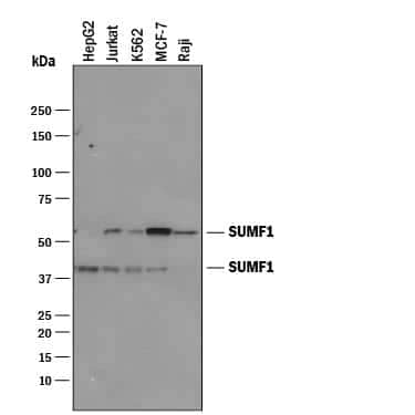
Species Human
Applications WB, ELISA, PA
|
|
![Western Blot: N-Acetylgalactosamine-6-Sulfatase/GALNS Antibody [NBP1-32899] - A. 30 ug PC-12 whole cell lysate/extract B. 30 ug Rat2 whole cell lysate/extract7.5% SDS-PAGE GALNS antibody dilution: 1:500. The HRP-conjugated anti-rabbit IgG antibody (NBP2-19301) was used to detect the primary antibody.](https://images.novusbio.com/fullsize/N-Acetylgalactosamine-6-Sulfatase-GALNS-Antibody-Western-Blot-NBP1-32899-img0012.jpg)
![Immunocytochemistry/Immunofluorescence: N-Acetylgalactosamine-6-Sulfatase/GALNS Antibody [NBP1-32899] - Paraformaldehyde-fixed HeLa, using GALNS antibody (Green) at 1:500 dilution. Alpha-tubulin filaments were labeled Red.](https://images.novusbio.com/fullsize/N-Acetylgalactosamine-6-Sulfatase-GALNS-Antibody-Immunocytochemistry-Immunofluorescence-NBP1-32899-img0006.jpg)
Rabbit Polyclonal
Species Human, Mouse, Rat
Applications WB, ICC/IF, IHC
| 2 Reviews |
|
![Western Blot: Aromatase Antibody [NB100-1596] - Expression of the cytochrome P450 aromatase in the ovary of control (C; open bar; n = 6) and hypothyroid (Hypo; solid bar; n = 6) rabbits. Representative immunoblot showing the expression of aromatase. Image collected and cropped by CiteAb from the following publication (https://www.hindawi.com/journals/bmri/2017/3795950/), licensed under a CC-BY license.](https://images.novusbio.com/fullsize/Aromatase-Antibody-Western-Blot-NB100-1596-img0005.jpg)
![Immunocytochemistry/Immunofluorescence: Aromatase Antibody [NB100-1596] - U2OS cells were fixed in 4% paraformaldehyde for 10 minutes and permeabilized in 0.05% Triton X-100 in PBS for 5 minutes. The cells were incubated with anti-Aromatase Antibody NB100-1596 at 2 ug/ml overnight at 4C and detected with an anti-rabbit Dylight 488 (Green) at a 1:1000 dilution for 60 minutes. Nuclei were counterstained with DAPI (Blue). Cells were imaged using a 100X objective and digitally deconvolved.](https://images.novusbio.com/fullsize/Aromatase-Antibody-Immunocytochemistry-Immunofluorescence-NB100-1596-img0008.jpg)
Rabbit Polyclonal
Species Human, Mouse, Rat
Applications WB, Simple Western, ICC/IF
| 16 Publications |
|
![Western Blot: DHPS Antibody [NBP1-82648] - Analysis in control (vector only transfected HEK293T lysate) and DHPS over-expression lysate (Co-expressed with a C-terminal myc-DDK tag (3.1 kDa) in mammalian HEK293T cells).](https://images.novusbio.com/fullsize/DHPS-Antibody-Western-Blot-NBP1-82648-img0008.jpg)
![Immunocytochemistry/Immunofluorescence: DHPS Antibody [NBP1-82648] - Staining of human cell line U-2 OS shows localization to nucleoplasm, plasma membrane & cytosol. Antibody staining is shown in green.](https://images.novusbio.com/fullsize/DHPS-Antibody-Immunocytochemistry-Immunofluorescence-NBP1-82648-img0007.jpg)
Rabbit Polyclonal
Species Human
Applications WB, ICC/IF, IHC
| 2 Publications |
|
![Western Blot: OXSM Antibody [NBP1-84732] - Lane 1: NIH-3T3 cell lysate (Mouse embryonic fibroblast cells) Lane 2: NBT-II cell lysate (Rat Wistar bladder tumour cells)](https://images.novusbio.com/fullsize/OXSM-Antibody-Western-Blot-NBP1-84732-img0006.jpg)
![Immunohistochemistry-Paraffin: OXSM Antibody [NBP1-84732] - Staining of human lymph node.](https://images.novusbio.com/fullsize/OXSM-Antibody-Immunohistochemistry-Paraffin-NBP1-84732-img0010.jpg)
Rabbit Polyclonal
Species Human, Mouse, Rat
Applications WB, IHC, IHC-P
|
|
![Western Blot: Peripherin 2/PRPH2 Antibody [NBP1-86687] - Analysis in control (vector only transfected HEK293T lysate) and PRPH2 over-expression lysate (Co-expressed with a C-terminal myc-DDK tag (3.1 kDa) in mammalian HEK293T cells).](https://images.novusbio.com/fullsize/Peripherin-2-PRPH2-Antibody-Western-Blot-NBP1-86687-img0006.jpg)
![Immunohistochemistry-Paraffin: Peripherin 2/PRPH2 Antibody [NBP1-86687] - Staining of human liver shows no positivity in hepatocytes as expected.](https://images.novusbio.com/fullsize/Peripherin-2-PRPH2-Antibody-Immunohistochemistry-Paraffin-NBP1-86687-img0010.jpg)
Rabbit Polyclonal
Species Human
Applications WB, IHC, IHC-P
|
|
![Immunohistochemistry-Paraffin: Dehydrodolichyl Diphosphate Synthase Antibody [NBP1-87964] - Staining of human Skeletal muscle shows very weak cytoplasmic positivity in myocytes.](https://images.novusbio.com/fullsize/Dehydrodolichyl-Diphosphate-Synthase-Antibody-Immunohistochemistry-Paraffin-NBP1-87964-img0006.jpg)
![Immunohistochemistry-Paraffin: Dehydrodolichyl Diphosphate Synthase Antibody [NBP1-87964] - Staining of human Epididymis shows strong membranous and cytoplasmic positivity in glandular cells.](https://images.novusbio.com/fullsize/Dehydrodolichyl-Diphosphate-Synthase-Antibody-Immunohistochemistry-Paraffin-NBP1-87964-img0003.jpg)
Rabbit Polyclonal
Species Human
Applications IHC, IHC-P
| 1 Publication |
|
![Immunohistochemistry-Paraffin: Steroid sulfatase Antibody [NBP1-90095] - Staining of human placenta shows high expression.](https://images.novusbio.com/fullsize/Steroid-sulfatase-Antibody-Immunohistochemistry-Paraffin-NBP1-90095-img0005.jpg)
![Immunohistochemistry-Paraffin: Steroid sulfatase Antibody [NBP1-90095] - Staining of human pancreas shows low expression as expected.](https://images.novusbio.com/fullsize/Steroid-sulfatase-Antibody-Immunohistochemistry-Paraffin-NBP1-90095-img0004.jpg)
Rabbit Polyclonal
Species Human
Applications IHC, IHC-P
| 3 Publications |
|
![Immunohistochemistry: POMC Antibody [NB100-1533] - Representative confocal images of POMC in POMC-transfected WT and Sel1L-/- N2a cells. White arrows point to POMC-containing secretory granules, while yellow arrows point to perinuclear POMC. KDEL marks the ER. Representative data from at least 2 independent experiments are shown. Image collected and cropped by CiteAb from the following publication (jci.org/articles/view/96420), licensed under a CC-BY license.](https://images.novusbio.com/fullsize/POMC-Antibody-Immunohistochemistry-NB100-1533-img0008.jpg)
![Flow Cytometry: POMC Antibody [NB100-1533] - Flow cytometric analysis of paraformaldehyde fixed A431 cells (blue line), permeabilized with 0.5% Triton. Primary incubation 1hr (10 ug/mL) followed by Alexa Fluor 488 secondary antibody (1 ug/mL). IgG control: Unimmunized goat IgG (black line) followed by Alexa Fluor 488 secondary antibody.](https://images.novusbio.com/fullsize/POMC-Antibody-Flow-Cytometry-NB100-1533-img0006.jpg)
Goat Polyclonal
Species Human, Mouse, Rat
Applications WB, Flow, ICC/IF
| 11 Publications |
|
Goat Polyclonal
Species Mouse
Applications WB, IP
|
|
Goat Polyclonal
Species Human
Applications WB
| 1 Publication |
|


Mouse Monoclonal
Species Human
Applications WB, ICC
| 1 Review 4 Publications |
|

Sheep Polyclonal
Species Human
Applications IHC
| 1 Publication |
|


Mouse Monoclonal
Species Human, Mouse
Applications WB, Simple Western, CyTOF-ready
| 3 Reviews 91 Publications |
|
