| Submit your blog on Female Sex Determination to be featured! |
| Submit your event on Female Sex Determination to be featured! |
![Western Blot: Aromatase Antibody [NB100-1596] - Expression of the cytochrome P450 aromatase in the ovary of control (C; open bar; n = 6) and hypothyroid (Hypo; solid bar; n = 6) rabbits. Representative immunoblot showing the expression of aromatase. Image collected and cropped by CiteAb from the following publication (https://www.hindawi.com/journals/bmri/2017/3795950/), licensed under a CC-BY license.](https://images.novusbio.com/fullsize/Aromatase-Antibody-Western-Blot-NB100-1596-img0005.jpg)
![Immunocytochemistry/Immunofluorescence: Aromatase Antibody [NB100-1596] - U2OS cells were fixed in 4% paraformaldehyde for 10 minutes and permeabilized in 0.05% Triton X-100 in PBS for 5 minutes. The cells were incubated with anti-Aromatase Antibody NB100-1596 at 2 ug/ml overnight at 4C and detected with an anti-rabbit Dylight 488 (Green) at a 1:1000 dilution for 60 minutes. Nuclei were counterstained with DAPI (Blue). Cells were imaged using a 100X objective and digitally deconvolved.](https://images.novusbio.com/fullsize/Aromatase-Antibody-Immunocytochemistry-Immunofluorescence-NB100-1596-img0008.jpg)
Rabbit Polyclonal
Species Human, Mouse, Rat
Applications WB, Simple Western, ICC/IF
| 16 Publications |
|
![Western Blot: SF-1/NR5A1/Steroidogenic Factor 1 Antibody [NBP1-52823] - Titration: 0.2-1 ug/ml, Positive Control: THP-1 cell lysate.](https://images.novusbio.com/fullsize/SF-1-NR5A1-Steroidogenic-Factor-1-Antibody-Western-Blot-NBP1-52823-img0007.jpg)
![Immunohistochemistry-Paraffin: SF-1/NR5A1/Steroidogenic Factor 1 Antibody [NBP1-52823] - Human adrenal tissue at an antibody concentration of 4-8ug/ml.](https://images.novusbio.com/fullsize/SF-1-NR5A1-Steroidogenic-Factor-1-Antibody-Immunohistochemistry-Paraffin-NBP1-52823-img0006.jpg)
Rabbit Polyclonal
Species Human, Mouse
Applications WB, Simple Western, ICC/IF
| 4 Publications |
|
![Immunohistochemistry-Paraffin: DMRT1 Antibody [NBP1-84071] - Staining in human testis and fallopian tube tissues using anti-DMRT1 antibody. Corresponding DMRT1 RNA-seq data are presented for the same tissues.](https://images.novusbio.com/fullsize/DMRT1-Antibody-Immunohistochemistry-Paraffin-NBP1-84071-img0010.jpg)
![Western Blot: DMRT1 Antibody [NBP1-84071] - Analysis in control (vector only transfected HEK293T lysate) and DMRT1 over-expression lysate (Co-expressed with a C-terminal myc-DDK tag (3.1 kDa) in mammalian HEK293T cells).](https://images.novusbio.com/fullsize/DMRT1-Antibody-Western-Blot-NBP1-84071-img0007.jpg)
Rabbit Polyclonal
Species Human
Applications WB, IHC, IHC-P
| 4 Publications |
|
![Immunohistochemistry-Paraffin: LYPLA3 Antibody [NBP1-92089] - Staining of human colon, kidney, lymph node and placenta using Anti-PLA2G15 antibody NBP1-92089 (A) shows similar protein distribution across tissues to independent antibody NBP1-92088 (B).](https://images.novusbio.com/fullsize/LYPLA3-Antibody-Immunohistochemistry-Paraffin-NBP1-92089-img0013.jpg)
![Western Blot: LYPLA3 Antibody [NBP1-92089] - Lane 1: Marker [kDa] 250, 130, 95, 72, 55, 36, 28, 17, 10. Lane 2: Human cell line RT-4. Lane 3: Human cell line U-251MG sp. Lane 4: Human plasma (IgG/HSA depleted). Lane 5: Human liver tissue. Lane 6: Human tonsil tissue](https://images.novusbio.com/fullsize/LYPLA3-Antibody-Western-Blot-NBP1-92089-img0004.jpg)
Rabbit Polyclonal
Species Human
Applications WB, IHC, IHC-P
|
|
![Western Blot: ZBTB22 Antibody [NBP1-92603] - Analysis in control (vector only transfected HEK293T lysate) and ZBTB22 over-expression lysate (Co-expressed with a C-terminal myc-DDK tag (3.1 kDa) in mammalian HEK293T cells).](https://images.novusbio.com/fullsize/ZBTB22-Antibody-Western-Blot-NBP1-92603-img0004.jpg)
![Immunocytochemistry/Immunofluorescence: ZBTB22 Antibody [NBP1-92603] - Staining of human cell line U-2 OS shows positivity in nucleus but not nucleoli.](https://images.novusbio.com/fullsize/ZBTB22-Antibody-Immunocytochemistry-Immunofluorescence-NBP1-92603-img0003.jpg)
Rabbit Polyclonal
Species Human
Applications WB, ICC/IF
| 1 Publication |
|
![Flow Cytometry: TCR alpha/beta Antibody (R73) [NBP2-12941] - Rat splenocytes with anti-rat TCR alpha/beta (R73) FITC.](https://images.novusbio.com/fullsize/TCR-alpha-beta-Antibody-R73-Flow-Cytometry-NBP2-12941-img0002.jpg)
![Flow Cytometry: TCR alpha/beta Antibody (R73) [NBP2-12941] - Rat thymocytes with anti-rat TCR alpha/beta (R73) PE.](https://images.novusbio.com/fullsize/TCR-alpha-beta-Antibody-R73-Flow-Cytometry-NBP2-12941-img0001.jpg)
Mouse Monoclonal
Species Rat, Primate
Applications WB, Flow, ICC/IF
| 1 Review 2 Publications |
|
![Western Blot: FOXL2 Antibody [NB100-1277] - (1ug/ml) staining of Mouse Ovary lysate (35ug protein in RIPA buffer). Detected by chemiluminescence.](https://images.novusbio.com/fullsize/antibody/nb100-1277_goat-polyclonal-foxl2-antibody-imgenex-img-3228-197202382424.jpg)
![Immunocytochemistry/Immunofluorescence: FOXL2 Antibody [NB100-1277] - Analysis of FOXL2 in PFA-fixed, E14.5 mouse ovary section using anti-FOXL2 antibody (magenta), germ cell marker (green) and hoechst (grey). ICC/IF image submitted by a verified customer review.](https://images.novusbio.com/fullsize/FOXL2-Antibody-Immunofluorescence-NB100-1277-img0020.jpg)
Goat Polyclonal
Species Human, Mouse, Bovine
Applications WB, ICC/IF, IHC
| 1 Review 72 Publications |
|


Goat Polyclonal
Species Human, Mouse
Applications WB, Simple Western, Flow
| 4 Reviews 110 Publications |
|


Goat Polyclonal
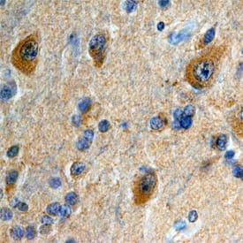
Species Human, Mouse, Rat
Applications WB, Simple Western, IHC
| 2 Reviews 31 Publications |
|


Goat Polyclonal
Species Human
Applications WB, Simple Western, ICC
| 2 Reviews 146 Publications |
|
Goat Polyclonal
Species Mouse
Applications WB, IHC
| 1 Review 12 Publications |
|

Sheep Polyclonal
Species Human
Applications WB
|
|
Sheep Polyclonal
Species Human
Applications IHC
| 1 Publication |
|
![Western Blot: SRY Antibody (1G4) [NBP2-37463] - Analysis using SRY mouse mAb against NTERA-2 (1) cell lysate.](https://images.novusbio.com/fullsize/SRY-Antibody-1G4-Western-Blot-NBP2-37463-img0003.jpg)
![Immunohistochemistry-Paraffin: SRY Antibody (1G4) [NBP2-37463] - Analysis of ovarian cancer tissues using SRY mouse mAb with DAB staining.](https://images.novusbio.com/fullsize/SRY-Antibody-1G4-Immunohistochemistry-NBP2-37463-img0006.jpg)
Mouse Monoclonal
Species Human
Applications WB, ELISA, Flow
|
|

Species Mouse
Applications BA
| 9 Publications |
|


Species Human
Applications BA
| 199 Publications |
|
![N/A Follistatin [HRP]](https://images.novusbio.com/fullsize2/DATA_Follistatin_DFN00_ELISA_681.jpg)
![N/A Follistatin [HRP]](https://images.novusbio.com/fullsize2/DATA_Follistatin_DFN00_ELISA_682.jpg)
Species Human
Applications ELISA
| 35 Publications |
|

Species Human, Mouse, Rat
Applications BA
| 187 Publications |
|
![Western Blot: RFC2 Antibody (OTI2B2) - Azide and BSA Free [NBP2-73857] - Analysis of HEK293T cells were transfected with the pCMV6-ENTRY control (Left lane) or pCMV6-ENTRY RFC2.](https://images.novusbio.com/fullsize/RFC2-Antibody-OTI2B2-Azide-and-BSA-Free-Western-Blot-NBP2-73857-img0001.jpg)
![Immunohistochemistry: RFC2 Antibody (OTI2B2) - Azide and BSA Free [NBP2-73857] - Analysis of Human prostate tissue.](https://images.novusbio.com/fullsize/RFC2-Antibody-OTI2B2-Azide-and-BSA-Free-Immunohistochemistry-NBP2-73857-img0012.jpg)
Mouse Monoclonal
Species Human, Mouse, Rat
Applications WB, Flow, IHC
|
|
