Disease and disorder research has been conducted in relation to the Cation Transport Pathway and Hypertensive Disease, Essential Hypertension, Edema, Retinoblastoma, Anemia. The study of the Cation Transport Pathway has been mentioned in research publications which can be found using our bioinformatics tool below. The Cation Transport Pathway has been researched in relation to Transport, Organic Cation Transport, Ion Transport, Secretion, Excretion. The Cation Transport Pathway complements our catalog of research reagents including antibodies and ELISA kits against RBC, ROCT1, AGT, ALB, BCHE.
Top Research Reagents
We have 2023 products for the study of the Cation Transport Pathway that can be applied to Flow Cytometry, Immunocytochemistry/ Immunofluorescence, Immunohistochemistry, Western Blot from our catalog of antibodies and ELISA kits.
![Western Blot: Serpin A8/Angiotensinogen Antibody [NBP1-30027] - Analysis of Angiotensinogen in human kidney lysate.](https://images.novusbio.com/fullsize/Serpin-A8-Angiotensinogen-Antibody-Western-Blot-NBP1-30027-img0013.jpg)
![Immunocytochemistry/Immunofluorescence: Serpin A8/Angiotensinogen Antibody [NBP1-30027] - HepG2 cells were fixed for 10 minutes using 10% formalin and then permeabilized for 5 minutes using 1X TBS + 0.5% Triton-X100. The cells were incubated with anti-Angiotensinogen at 10 ug/ml overnight at 4C and detected with an anti-rabbit Dylight 488 (Green) at a 1:500 dilution. Alpha tubulin (DM1A) NB100-690 was used as a co-stain at a 1:1000 dilution and detected with an anti-mouse Dylight 550 (Red) at a 1:500 dilution. Nuclei were counterstained with DAPI (Blue). Cells were imaged using a 40X objective.](https://images.novusbio.com/fullsize/Serpin-A8-Angiotensinogen-Antibody-Immunocytochemistry-Immunofluorescence-NBP1-30027-img0014.jpg)
![Western Blot: SLC22A1 Antibody (2C5) [NBP1-51684] - Analysis using SLC22A1 mAb against HEK293 (1) and SLC22A1(AA: 284-347)-hIgGFc transfected HEK293 (2) cell lysate.](https://images.novusbio.com/fullsize/SLC22A1-Antibody-2C5-Western-Blot-NBP1-51684-img0007.jpg)
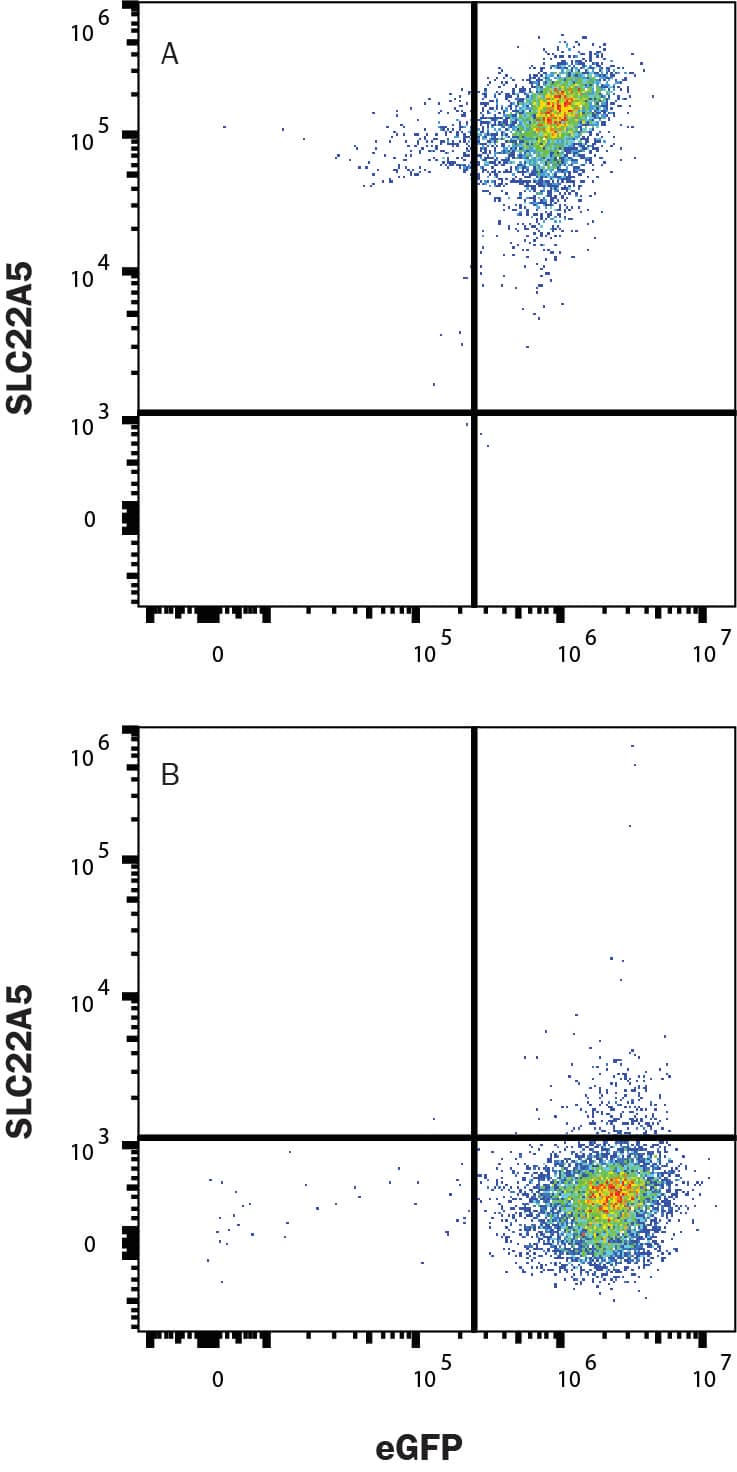
![Flow Cytometry: SLC22A1 Antibody (2C5) [NBP1-51684] - Analysis of Jurkat cells using SLC22A1 mouse mAb (green) and negative control (purple).](https://images.novusbio.com/fullsize/SLC22A1-Antibody-2C5-Flow-Cytometry-NBP1-51684-img0005.jpg)
![Immunohistochemistry-Paraffin: DBT Antibody [NBP1-89522] - Analysis in human kidney and pancreas tissues. Corresponding DBT RNA-seq data are presented for the same tissues.](https://images.novusbio.com/fullsize/DBT-Antibody-Immunohistochemistry-Paraffin-NBP1-89522-img0012.jpg)
![Immunohistochemistry-Paraffin: DBT Antibody [NBP1-89522] - Staining of human colon, liver, lymph node and pancreas using Anti-DBT antibody NBP1-89522 (A) shows similar protein distribution across tissues to independent antibody NBP1-85964 (B).](https://images.novusbio.com/fullsize/DBT-Antibody-Immunohistochemistry-Paraffin-NBP1-89522-img0013.jpg)
![Western Blot: MASA Antibody [NBP2-13961] - Analysis in control (vector only transfected HEK293T lysate) and ENOPH1 over-expression lysate (Co-expressed with a C-terminal myc-DDK tag (3.1 kDa) in mammalian HEK293T cells).](https://images.novusbio.com/fullsize/MASA-Antibody-Western-Blot-NBP2-13961-img0009.jpg)
![Immunocytochemistry/Immunofluorescence: MASA Antibody [NBP2-13961] - Staining of human cell line MCF7 shows localization to nucleoplasm & nuclear bodies.](https://images.novusbio.com/fullsize/MASA-Antibody-Immunocytochemistry-Immunofluorescence-NBP2-13961-img0005.jpg)
![Western Blot: OCT1 Antibody [NBP2-21584] - Various whole cell extracts (30 ug) were separated by 7.5% SDS-PAGE, and the membrane was blotted with OCT1 antibody diluted at 1:1000.](https://images.novusbio.com/fullsize/OCT1-Antibody-Western-Blot-NBP2-21584-img0011.jpg)
![Immunocytochemistry/Immunofluorescence: OCT1 Antibody [NBP2-21584] - HeLa cells were fixed in 4% paraformaldehyde at RT for 15 min. Green: OCT1 protein stained by OCT1 antibody diluted at 1:500. Red: phalloidin, a cytoskeleton marker, stained by () diluted at 1:200. Blue: Hoechst 33342 staining. Scale bar = 10 um.](https://images.novusbio.com/fullsize/OCT1-Antibody-Immunocytochemistry-Immunofluorescence-NBP2-21584-img0006.jpg)


![Western Blot: SLC29A4 Antibody [NBP2-41314] - Western blot analysis of SLC29A4 in SK-N-SH cell lysate with SLC29A4 antibody at 1 ug/ml.](https://images.novusbio.com/fullsize/SLC29A4-Antibody-Western-Blot-NBP2-41314-img0001.jpg)
![Immunocytochemistry/ Immunofluorescence: SLC29A4 Antibody - BSA Free [NBP2-41314] - Immunofluorescence of SLC29A4 in human brain tissue with SLC29A4 antibody at 20 u/mL.](https://images.novusbio.com/fullsize/nbp2-41314_rabbit-polyclonal-slc29a4-antibody-271120242004191.jpg)


![Immunohistochemistry-Paraffin: Phenylalanine Hydroxylase Antibody [NBP2-48615] - Analysis in human liver and pancreas tissues. Corresponding Phenylalanine Hydroxylase RNA-seq data are presented for the same tissues.](https://images.novusbio.com/fullsize/Phenylalanine-Hydroxylase-Antibody-Immunohistochemistry-Paraffin-NBP2-48615-img0012.jpg)
![Immunohistochemistry-Paraffin: Phenylalanine Hydroxylase Antibody [NBP2-48615] - Staining of human cerebral cortex, kidney, liver and pancreas using Anti-Phenylalanine Hydroxylase antibody NBP2-48615 (A) shows similar protein distribution across tissues to independent antibody NBP1-80917 (B).](https://images.novusbio.com/fullsize/Phenylalanine-Hydroxylase-Antibody-Immunohistochemistry-Paraffin-NBP2-48615-img0014.jpg)
![Immunohistochemistry-Paraffin: Band 3 Antibody [NBP2-49409] - Staining of human cerebral cortex shows low expression as expected.](https://images.novusbio.com/fullsize/Band-3-Antibody-Immunohistochemistry-Paraffin-NBP2-49409-img0003.jpg)
![Immunohistochemistry: Band 3 Antibody [NBP2-49409] - Staining of human spleen shows strong positivity in erythrocytes.](https://images.novusbio.com/fullsize/Band-3-Antibody-Immunohistochemistry-NBP2-49409-img0001.jpg)
![Western Blot: POU2F2 Antibody (Oct2/2137) [NBP2-75734] - Analysis (A) Recombinant Protein (B) Daudi cell lysate POU2F2 Mouse Monoclonal Antibody (OCT2/2137).](https://images.novusbio.com/fullsize/OCT2-Antibody-Oct2-2137-Western-Blot-NBP2-75734-img0004.jpg)
![Immunohistochemistry-Paraffin: POU2F2 Antibody (Oct2/2137) [NBP2-75734] - Formalin-fixed, paraffin-embedded human Tonsil stained with POU2F2 Mouse Monoclonal Antibody (OCT2/2137).](https://images.novusbio.com/fullsize/OCT2-Antibody-Oct2-2137-Immunohistochemistry-Paraffin-NBP2-75734-img0002.jpg)
