| Submit your blog on Plagiocephaly to be featured! |
| Submit your event on Plagiocephaly to be featured! |
Mouse Monoclonal
Applications IHC
|
|
![Western Blot: Inorganic Pyrophosphatase/PPA1 Antibody [NBP1-31348] - A. 30 ug Neuro2A whole cell lysate/extract. B. 30 ug GL261 whole cell lysate/extract. C. 30 ug C8D30 whole cell lysate/extract. D. 30 ug NIH-3T3 whole cell lysate/extract. E. 30 ug BCL-1 whole cell lysate/extract. F. 30 ug Raw264.7 whole cell lysate/extract. G. 30 ug C2C12 whole cell lysate/extract.](https://images.novusbio.com/fullsize/Inorganic-Pyrophosphatase-PPA1-Antibody-Western-Blot-NBP1-31348-img0012.jpg)
![Immunocytochemistry/Immunofluorescence: Inorganic Pyrophosphatase/PPA1 Antibody [NBP1-31348] - Paraformaldehyde-fixed HeLa, using antibody at 1:200 dilution.](https://images.novusbio.com/fullsize/Inorganic-Pyrophosphatase-PPA1-Antibody-Immunocytochemistry-Immunofluorescence-NBP1-31348-img0008.jpg)
Rabbit Polyclonal
Species Human, Mouse, Rat
Applications WB, ICC/IF, IHC
|
|
![Western Blot: SRPR alpha Antibody [H00006734-B02P] - Analysis of SRPR expression in human pancreas.](https://images.novusbio.com/fullsize/SRPR-alpha-Antibody-Western-Blot-H00006734-B02P-img0002.jpg)
![Immunocytochemistry/Immunofluorescence: SRPR alpha Antibody [H00006734-B02P] - Analysis of purified antibody to SRPR on HeLa cell. (antibody concentration 10 ug/ml)](https://images.novusbio.com/fullsize/SRPR-alpha-Antibody-Immunocytochemistry-Immunofluorescence-H00006734-B02P-img0001.jpg)
Mouse Polyclonal
Species Human
Applications WB, ICC/IF
| 1 Publication |
|
![Immunohistochemistry-Paraffin: CHM Antibody [NBP1-84954] - Staining of human cerebral cortex shows moderate positivity in neurons.](https://images.novusbio.com/fullsize/CHM-Antibody-Immunohistochemistry-Paraffin-NBP1-84954-img0007.jpg)
![Immunohistochemistry-Paraffin: CHM Antibody [NBP1-84954] - Staining of human cerebellum shows strong positivity in cells in molecular layer.](https://images.novusbio.com/fullsize/CHM-Antibody-Immunohistochemistry-Paraffin-NBP1-84954-img0006.jpg)
Rabbit Polyclonal
Species Human
Applications IHC, IHC-P
|
|
![Western Blot: NDUFB6 Antibody [NBP1-92172] - Lane 1: Marker [kDa] 250, 130, 95, 72, 55, 36, 28, 17, 10. Lane 2: Human cell line RT-4. Lane 3: Human cell line U-251MG sp. Lane 4: Human plasma (IgG/HSA depleted). Lane 5: Human liver tissue. Lane 6: Human tonsil tissue](https://images.novusbio.com/fullsize/NDUFB6-Antibody-Western-Blot-NBP1-92172-img0006.jpg)
![Immunocytochemistry/Immunofluorescence: NDUFB6 Antibody [NBP1-92172] - Immunofluorescent staining of human cell line A-431 shows localization to nucleoplasm & mitochondria.](https://images.novusbio.com/fullsize/NDUFB6-Antibody-Immunocytochemistry-Immunofluorescence-NBP1-92172-img0007.jpg)
Rabbit Polyclonal
Species Human
Applications WB, ICC/IF, IHC
| 2 Publications |
|
![Western Blot: RanGAP1 Antibody (1B4) [NBP2-02623] Analysis of extracts (35ug) from 9 different cell lines by using anti-RanGAP1 monoclonal antibody.](https://images.novusbio.com/fullsize/RanGAP1-Antibody-1B4-Western-Blot-NBP2-02623-img0009.jpg)
![Immunocytochemistry/Immunofluorescence: RanGAP1 Antibody (1B4) [NBP2-02623] - Staining of COS7 cells transiently transfected by pCMV6-ENTRY RanGAP1.](https://images.novusbio.com/fullsize/RanGAP1-Antibody-1B4-Immunocytochemistry-Immunofluorescence-NBP2-02623-img0008.jpg)
Mouse Monoclonal
Species Human, Mouse, Rat
Applications WB, Flow, ICC/IF
| 1 Publication |
|
![Western Blot: SLC17A5 Antibody [NBP2-20383] - Sample (30 ug of whole cell lysate) A: Jurkat 10% SDS PAGE gel, diluted at 1:1000.](https://images.novusbio.com/fullsize/SLC17A5-Antibody-Western-Blot-NBP2-20383-img0001.jpg)
![Immunocytochemistry/Immunofluorescence: SLC17A5 Antibody [NBP2-20383] - Sample: HepG2 cells were fixed in -20C 100% MeOH for 5 min. Green: SLC17A5 protein stained by SLC17A5 antibody diluted at 1:500. Blue: Hoechst 33343 staining.](https://images.novusbio.com/fullsize/SLC17A5-Antibody-Immunocytochemistry-Immunofluorescence-NBP2-20383-img0003.jpg)
Rabbit Polyclonal
Species Human, Mouse
Applications WB, ICC/IF, IHC
|
|
![Western Blot: Twist-1 Antibody (10E4E6) [NBP2-37364] - Analysis using TWIST1 mouse mAb against NIH/3T3 (1), JURKAT (2), HELA (3), A549 (4), RAJI (5) and OCM-1 (6) cell lysate.](https://images.novusbio.com/fullsize/Twist-1-Antibody-10E4E6-Western-Blot-NBP2-37364-img0004.jpg)
![Immunocytochemistry/Immunofluorescence: Twist-1 Antibody (10E4E6) [NBP2-37364] - Analysis of HeLa cells using TWIST1 mouse mAb (green). Blue: DRAQ5 fluorescent DNA dye.](https://images.novusbio.com/fullsize/Twist-1-Antibody-10E4E6-Immunocytochemistry-NBP2-37364-img0011.jpg)
Mouse Monoclonal
Species Human, Mouse
Applications WB, ELISA, Flow
| 1 Review 9 Publications |
|

Species Human
Applications BA
| 3 Publications |
|


Mouse Monoclonal
Species Human
Applications WB, Flow, IHC
| 1 Review 3 Publications |
|
![Immunohistochemistry-Paraffin: LAMC2 Antibody (CL2980) [NBP2-42388] - Staining in human fallopian tube and liver tissues. Corresponding LAMC2 RNA-seq data are presented for the same tissues.](https://images.novusbio.com/fullsize/LAMC2-Antibody-CL2980-Immunohistochemistry-Paraffin-NBP2-42388-img0023.jpg)
![Western Blot: LAMC2 Antibody (CL2980) [NBP2-42388] - Analysis in A-431 cells transfected with control siRNA, target specific siRNA probe #1 and #2, using Anti-LAMC2 antibody. Remaining relative intensity is presented. Loading control: Anti-GAPDH.](https://images.novusbio.com/fullsize/LAMC2-Antibody-CL2980-Western-Blot-NBP2-42388-img0017.jpg)
Mouse Monoclonal
Species Human
Applications WB, ICC/IF, IHC
| 4 Publications |
|
![Immunohistochemistry-Paraffin: Desmoplakin Antibody [NBP2-48836] - Staining in human skin and skeletal muscle tissues using anti-DSP antibody. Corresponding DSP RNA-seq data are presented for the same tissues.](https://images.novusbio.com/fullsize/Desmoplakin-Antibody-Immunohistochemistry-Paraffin-NBP2-48836-img0005.jpg)
![Immunohistochemistry-Paraffin: Desmoplakin Antibody [NBP2-48836] - Staining of human skeletal muscle shows low expression as expected.](https://images.novusbio.com/fullsize/Desmoplakin-Antibody-Immunohistochemistry-Paraffin-NBP2-48836-img0003.jpg)
Rabbit Polyclonal
Species Human, Canine
Applications ICC/IF, IHC, IHC-P
| 2 Publications |
|
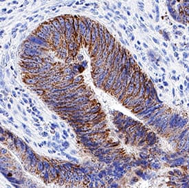
![Immunohistochemistry: Blood Group Lewis b Antibody (2-25LE) [NB500-526] - Immunohistochemistry staining of human colon adenocarcinoma (paraffin sections) using anti-blood group Lewis b (clone 2-25LE).](https://images.novusbio.com/fullsize/Blood-Group-Lewis-b-Antibody-2-25LE-Immunohistochemistry-NB500-526-img0002.jpg)
![Immunohistochemistry: Blood Group Lewis b Antibody (2-25LE) [NB500-526] - Staining of human small intestine (paraffin sections) using anti-blood group Lewis b (clone 2-25LE).](https://images.novusbio.com/fullsize/Blood-Group-Lewis-b-Antibody-2-25LE-Immunohistochemistry-NB500-526-img0001.jpg)
Mouse Monoclonal
Species Human
Applications WB, ELISA, IHC
| 3 Publications |
|
![Western Blot: PRRT2 Antibody [NBP2-82026] - Analysis of PRRT2 in mouse brain tissue lysate with PRRT2 antibody at 1 ug/ml.](https://images.novusbio.com/fullsize/PRRT2-Antibody-Western-Blot-NBP2-82026-img0001.jpg)
![Immunocytochemistry/ Immunofluorescence: PRRT2 Antibody - BSA Free [NBP2-82026] - Immunofluorescence of PRRT2 in rat brain tissue with PRRT2 antibody at 20 u/ml.](https://images.novusbio.com/fullsize/nbp2-82026_rabbit-polyclonal-prrt2-antibody-2711202419172250.jpg)
Rabbit Polyclonal
Species Human, Mouse, Rat
Applications WB, ELISA, ICC/IF
|
|
![Immunocytochemistry/Immunofluorescence: NSP 5 alpha 3 alpha Antibody [NB100-517] - Immunostaining of HN30 and MCF-12a cells. MCF-12a NT (DAPI): non-treated MCF-12a DAPI stained, MCF-12a NT (NSP 5a3a-FITC): non-treated MCF-12a FITC stained for NSP 5a3a, MCF-12a pNSP 5a3a (DAPI): HN30 NT (DAPI): non-treated HN30 DAPI stained, HN30 NT (NSP 5a3a-FITC): non-treated HN30 FITC stained for NSP 5a3a, HN30 pNSP 5a3a (DAPI): White arrows indicate apoptotic bodies. Image collected and cropped by CiteAb from the following publication (https://www.oncotarget.com/article/306/text/) licensed under a CC-BY license.](https://images.novusbio.com/fullsize/NSP-5-alpha-3-alpha-Antibody-Immunocytochemistry-Immunofluorescence-NB100-517-img0005.jpg)
![Western Blot: NSP 5 alpha 3 alpha Antibody [NB100-517] - Western Blot analysis of total lysates from asynchronous HN30 cells and MCF-12a 3 days post-transfection. NT: non-treated, OB: only buffer, EGFP: only pcDNA3.1/CT-GFP vector, NSP 5a3a/EGFP: pcDNA 3.1/CT-GFP and pcDNA 3.0 NSP 5a3a. Image collected and cropped by CiteAb from the following publication (https://www.oncotarget.com/article/306/text/) licensed under a CC-BY license.](https://images.novusbio.com/fullsize/NSP-5-alpha-3-alpha-Antibody-Western-Blot-NB100-517-img0007.jpg)
Rabbit Polyclonal
Species Human
Applications WB, ICC/IF, IP
| 2 Publications |
|
![Western Blot: FGFR2 Antibody (1G3) [H00002263-M01] - Analysis of FGFR2 expression in transfected 293T cell line by FGFR2 monoclonal antibody (M01), clone 1G3.Lane 1: FGFR2 transfected lysate (Predicted MW: 92 KDa).Lane 2: Non-transfected lysate.](https://images.novusbio.com/fullsize/FGFR2-Antibody-1G3-Western-Blot-H00002263-M01-img0008.jpg)
![Immunohistochemistry-Paraffin: FGFR2 Antibody (1G3) [H00002263-M01] - Analysis of monoclonal antibody to FGFR2 on formalin-fixed paraffin-embedded human stomach carcinoma tissue. Antibody concentration 5 ug/ml.](https://images.novusbio.com/fullsize/FGFR2-Antibody-1G3-Immunohistochemistry-Paraffin-H00002263-M01-img0006.jpg)
Mouse Monoclonal
Species Human, Bovine
Applications WB, ELISA, ICC/IF
| 13 Publications |
|
![Western Blot: Adenine Nucleotide Translocator 2 Antibody [NBP2-92630] - Analysis of extracts of various cell lines, using Adenine Nucleotide Translocator 2 at 1:1000 dilution. Secondary antibody: HRP Goat Anti-Rabbit IgG (H+L) at 1:10000 dilution. Lysates/proteins: 25ug per lane. Blocking buffer: 3% nonfat dry milk in TBST. Detection: ECL Basic Kit . Exposure time: 60s.](https://images.novusbio.com/fullsize/Adenine-Nucleotide-Translocator-2-Antibody-Western-Blot-NBP2-92630-img0004.jpg)
![Immunocytochemistry/Immunofluorescence: Adenine Nucleotide Translocator 2 Antibody [NBP2-92630] - Analysis of HeLa cells using Adenine Nucleotide Translocator 2 . Blue: DAPI for nuclear staining.](https://images.novusbio.com/fullsize/Adenine-Nucleotide-Translocator-2-Antibody-Immunocytochemistry-Immunofluorescence-NBP2-92630-img0001.jpg)
Rabbit Polyclonal
Species Human, Mouse, Rat
Applications WB, ELISA, ICC/IF
|
|
