| Submit your blog on Lesion Of Ulnar Nerve to be featured! |
| Submit your event on Lesion Of Ulnar Nerve to be featured! |
![Western Blot: VAChT/SLC18A3 Antibody [NB100-91348] - Rat and mouse tissue lysates. Blocking: 1% LFDM for 30 min at RT; primary antibody: dilution 1:2000 incubated at 4C overnight.](https://images.novusbio.com/fullsize/VAChT-SLC18A3-Antibody-Western-Blot-NB100-91348-img0032.jpg)
![Immunohistochemistry-Paraffin: VAChT/SLC18A3 Antibody [NB100-91348] - Rat spinal cord. The animal was perfused at a pressure of 130 mmHg with 300 ml 4% FA before being processed for paraffin embedding. HIER: Tris-EDTA, pH 9 for 20 min. Blocking: 0.2% LFDM in TBST filtered thru 0.2 um.](https://images.novusbio.com/fullsize/VAChT-SLC18A3-Antibody-Immunohistochemistry-Paraffin-NB100-91348-img0031.jpg)
Rabbit Polyclonal
Species Human, Mouse, Rat
Applications WB, IHC, IHC-P
| 2 Publications |
|
![Western Blot: UVRAG Antibody [NBP1-18885] - Beclin 1 is acetylated at lysines 430 and 437. TSA and NAM increase the binding of Beclin 1 to Rubicon. Immunoprecipitation of indicated Beclin 1-binding partners with ectopically expressed Flag-Beclin 1 in HEK293T cells treated with TSA and NAM. Image collected and cropped by CiteAb from the following publication (https://www.nature.com/articles/ncomms8215), licensed under a CC-BY license.](https://images.novusbio.com/fullsize/UVRAG-Antibody-Western-Blot-NBP1-18885-img0005.jpg)
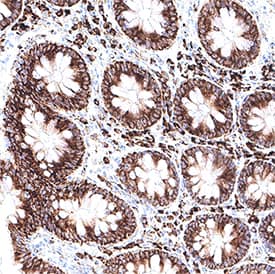
![Immunohistochemistry-Paraffin: UVRAG Antibody [NBP1-18885] - Section of human colon carcinoma. Antibody: Affinity purified rabbit anti- UVRAG used at a dilution of 1:200 (1ug/ml). Detection: DAB](https://images.novusbio.com/fullsize/UVRAG-Antibody-Immunohistochemistry-Paraffin-NBP1-18885-img0004.jpg)
Rabbit Polyclonal
Species Human
Applications WB, ICC/IF, IHC
| 4 Publications |
|
Mouse Monoclonal
Species Human
Applications WB, Simple Western, IHC
|
|
![Immunocytochemistry/Immunofluorescence: RPS3 Antibody [NBP1-33691] - Analysis of methanol-fixed HeLa, using RPS3 antibody (Green) at 1:500 dilution. Alpha-tubulin filaments were labeled with an alpha Tubulin antibody (Red) at 1:2000.](https://images.novusbio.com/fullsize/RPS3-Antibody-Immunocytochemistry-Immunofluorescence-NBP1-33691-img0003.jpg)
![Immunohistochemistry-Paraffin: RPS3 Antibody [NBP1-33691] - Mouse intestine diluted at 1:500. Antigen Retrieval: Citrate buffer, pH 6.0, 15 min.](https://images.novusbio.com/fullsize/RPS3-Antibody-Immunohistochemistry-Paraffin-NBP1-33691-img0009.jpg)
Rabbit Polyclonal
Species Human, Mouse, Rat
Applications WB, ICC/IF, IHC
|
|
![Western Blot: GARS Antibody [NBP1-85533] - Analysis using Anti-GARS antibody NBP1-85533 (A) shows similar pattern to independent antibody NBP1-85534 (B).](https://images.novusbio.com/fullsize/GARS-Antibody-Western-Blot-NBP1-85533-img0008.jpg)
![Immunocytochemistry/Immunofluorescence: GARS Antibody [NBP1-85533] - Immunofluorescent staining of human cell line U-2 OS shows localization to cytosol. Antibody staining is shown in green.](https://images.novusbio.com/fullsize/GARS-Antibody-Immunocytochemistry-Immunofluorescence-NBP1-85533-img0006.jpg)
Rabbit Polyclonal
Species Human
Applications WB, ICC/IF, IHC
|
|
![Western Blot: HIP-55 Antibody [NBP1-91988] - Analysis using Anti-DBNL antibody NBP1-91988 (A) shows similar pattern to independent antibody NBP1-91989 (B).](https://images.novusbio.com/fullsize/HIP-55-Antibody-Western-Blot-NBP1-91988-img0011.jpg)
![Immunohistochemistry-Paraffin: HIP-55 Antibody [NBP1-91988] - Staining of human testis.](https://images.novusbio.com/fullsize/HIP-55-Antibody-Immunohistochemistry-Paraffin-NBP1-91988-img0013.jpg)
Rabbit Polyclonal
Species Human, Mouse, Rat
Applications WB, IHC, IHC-P
|
|
![Western Blot: PSMD10 Antibody (3F6) [NBP2-02199] Analysis of extracts (35ug) from 9 different cell lines by using anti-PSMD10 monoclonal antibody.](https://images.novusbio.com/fullsize/PSMD10-Antibody-3F6-Western-Blot-NBP2-02199-img0015.jpg)
![Immunohistochemistry-Paraffin: PSMD10 Antibody (3F6) [NBP2-02199] - Staining of paraffin-embedded Ovary tissue using anti-PSMD10 mouse monoclonal antibody.](https://images.novusbio.com/fullsize/PSMD10-Antibody-3F6-Immunohistochemistry-Paraffin-NBP2-02199-img0013.jpg)
Mouse Monoclonal
Species Human, Mouse, Rat
Applications WB, Flow, IHC
| 1 Publication |
|
![Immunohistochemistry-Paraffin: Cystatin F Antibody [NBP2-13881] - Staining in human bone marrow and skeletal muscle tissues using NBP2-13881 antibody. Corresponding CST7 RNA-seq data are presented for the same tissues.](https://images.novusbio.com/fullsize/Cystatin-F-Antibody-Immunohistochemistry-Paraffin-NBP2-13881-img0013.jpg)
![Immunohistochemistry-Paraffin: Cystatin F Antibody [NBP2-13881] - Staining of human bone marrow shows high expression.](https://images.novusbio.com/fullsize/Cystatin-F-Antibody-Immunohistochemistry-Paraffin-NBP2-13881-img0007.jpg)
Rabbit Polyclonal
Species Human
Applications IHC, IHC-P
| 2 Reviews |
|
![Immunocytochemistry/Immunofluorescence: RPLP2 Antibody [NBP2-15104] - Analysis of methanol-fixed A431, using antibody at 1:500 dilution.](https://images.novusbio.com/fullsize/RPLP2-Antibody-Immunocytochemistry-Immunofluorescence-NBP2-15104-img0001.jpg)
![Immunohistochemistry-Paraffin: RPLP2 Antibody [NBP2-15104] - Human colon carcinoma, using RPLP2 antibody at 1:500 dilution. Antigen Retrieval: Trilogy&#8482; (EDTA based, pH 8.0) buffer, 15min.](https://images.novusbio.com/fullsize/RPLP2-Antibody-Immunohistochemistry-Paraffin-NBP2-15104-img0002.jpg)
Rabbit Polyclonal
Species Human
Applications WB, ICC/IF, IHC
|
|
![Immunocytochemistry/Immunofluorescence: DNALI1 Antibody [NBP2-16197] - Immunofluorescence analysis of methanol-fixed A549, using antibody at 1:500 dilution.](https://images.novusbio.com/fullsize/DNALI1-Antibody-Immunocytochemistry-Immunofluorescence-NBP2-16197-img0002.jpg)
![Immunohistochemistry-Paraffin: DNALI1 Antibody [NBP2-16197] - Paraffin-embedded mouse muscle. DNALI1 antibody diluted at 1:500.](https://images.novusbio.com/fullsize/DNALI1-Antibody-Immunohistochemistry-Paraffin-NBP2-16197-img0005.jpg)
Rabbit Polyclonal
Species Human, Mouse
Applications WB, ICC/IF, IHC
|
|
![Western Blot: GART Antibody (4D6-1D5) [H00002618-M01] - Blot showing the effect of hypoxia on the protein expression levels of the purine biosynthetic enzymes. HIF-1 alpha is stabilized in hypoxia as expected, and no significant increase in the purine enzymes was detected between normoxic (21% oxygen) and hypoxic (1% oxygen) growth conditions. The positions of molecular markers surrounding each band of interest are shown for each blot. ADSL (NBP2-03107), ATIC (NBP2-01941), FGAMS (NBP1-84691), GART (H00002618-M01), HIF-1a (NB100-449), PAICS (NBP2-02817), PPAT (NBP2-02056). Image collected and cropped by CiteAb from the following publication (//pubmed.ncbi.nlm.nih.gov/32439803/) licensed under a CC-BY license.](https://images.novusbio.com/fullsize/GART-Antibody-4D6-1D5-Western-Blot-H00002618-M01-img0012.jpg)
![Immunocytochemistry/Immunofluorescence: GART Antibody (4D6-1D5) [H00002618-M01] - Analysis of monoclonal antibody to GART on HeLa cell. Antibody concentration 10 ug/ml.](https://images.novusbio.com/fullsize/GART-Antibody-4D6-1D5-Immunocytochemistry-Immunofluorescence-H00002618-M01-img0006.jpg)
Mouse Monoclonal
Species Human, Mouse
Applications WB, ELISA, ICC/IF
| 9 Publications |
|


Sheep Polyclonal
Species Human
Applications WB, IHC
|
|

Goat Polyclonal
Species Human
Applications IHC
| 1 Publication |
|
Goat Polyclonal
Species Human
Applications WB, IHC
| 6 Publications |
|

Mouse Monoclonal
Species Human, Mouse, Rat
Applications WB
| 1 Publication |
|
![Western Blot: GRIP1 Antibody [NBP2-41211] - Analysis of GRIP1 in 293 cell lysate with GRIP1 antibody at 1 ug/mL in (A) the absence and (B) the presence of blocking peptide.](https://images.novusbio.com/fullsize/GRIP1-Antibody-Western-Blot-NBP2-41211-img0004.jpg)
![Immunohistochemistry: GRIP1 Antibody [NBP2-41211] - Immunohistochemistry of GRIP1 in rat brain tissue with GRIP1 antibody at 2.5 ug/ml.](https://images.novusbio.com/fullsize/GRIP1-Antibody-Immunohistochemistry-NBP2-41211-img0002.jpg)
Rabbit Polyclonal
Species Human, Mouse, Rat
Applications WB, ELISA, ICC/IF
|
|
![Western Blot: Acetylcholinesterase/ACHE Antibody [NB100-1519] - Staining (0.3ug/ml) of Jurkat (A) and (0.5ug/ml) HepG2 (B) cell lysate (35ug protein in RIPA buffer). Detected by chemiluminescence.](https://images.novusbio.com/fullsize/Acetylcholinesterase-ACHE-Antibody-Western-Blot-NB100-1519-img0006.jpg)
![Immunocytochemistry/Immunofluorescence: Acetylcholinesterase/ACHE Antibody [NB100-1519] - Immunofluorescence analysis of paraformaldehyde fixed U2OS cells, permeabilized with 0.15% Triton. Primary incubation 1hr (10ug/ml) followed by Alexa Fluor 488 secondary antibody (2ug/ml), showing nuclear, membrane and cytoplasmic staining. The nuclear stain is DAPI (blue). Negative control: Unimmunized goat IgG (10ug/ml) followed by Alexa Fluor 488 secondary antibody (2ug/ml).](https://images.novusbio.com/fullsize/Acetylcholinesterase-ACHE-Antibody-Immunocytochemistry-Immunofluorescence-NB100-1519-img0005.jpg)
Goat Polyclonal
Species Human, Mouse, Rat
Applications WB, Flow, ICC/IF
| 1 Review 1 Publication |
|
![Western Blot: Transthyretin/Prealbumin Antibody (2E10C5) [NBP2-52575] - Analysis using TTR mAb against human TTR (AA: 1-147) recombinant protein. (Expected MW is 45.8 kDa)](https://images.novusbio.com/fullsize/Transthyretin-Prealbumin-Antibody-2E10C5-Western-Blot-NBP2-52575-img0005.jpg)
![Immunocytochemistry/Immunofluorescence: Transthyretin/Prealbumin Antibody (2E10C5) [NBP2-52575] - Analysis of MCF-7 cells using TTR mouse mAb (green). Blue: DRAQ5 fluorescent DNA dye. Red: Actin filaments have been labeled with Alexa Fluor- 555 phalloidin.](https://images.novusbio.com/fullsize/Transthyretin-Prealbumin-Antibody-2E10C5-Immunofluorescence-NBP2-52575-img0003.jpg)
Mouse Monoclonal
Species Human
Applications WB, ELISA, Flow
| 1 Review 1 Publication |
|
![Western Blot: Proteasome 19S S3 Antibody (OTI2A2) - Azide and BSA Free [NBP2-73636] - HEK293T cells were transfected with the pCMV6-ENTRY control (Left lane) or pCMV6-ENTRY Proteasome 19S S3 (Right lane) cDNA for 48 hrs and lysed. Equivalent amounts of cell lysates (5 ug per lane) were separated by SDS-PAGE and immunoblotted with anti-Prot](https://images.novusbio.com/fullsize/Proteasome-19S-S3-Antibody-OTI2A2-Azide-and-BSA-Free-Western-Blot-NBP2-73636-img0002.jpg)
![Flow Cytometry: Proteasome 19S S3 Antibody (OTI2A2) - Azide and BSA Free [NBP2-73636] - HEK293T cells transfected with either overexpression plasmid (Red) or empty vector control plasmid (Blue) were immunostaining by anti-Proteasome 19S S3 antibody, and then analyzed by flow cytometry.](https://images.novusbio.com/fullsize/Proteasome-19S-S3-Antibody-OTI2A2-Azide-and-BSA-Free-Flow-Cytometry-NBP2-73636-img0005.jpg)
Mouse Monoclonal
Species Human, Mouse, Rat
Applications WB, Flow, CyTOF-ready
|
|
