| Submit your blog on Keratosis Follicularis to be featured! |
| Submit your event on Keratosis Follicularis to be featured! |
Mouse Monoclonal
Applications IHC
|
|
![Western Blot: SERCA3 ATPase Antibody (2H3) [H00000489-M01] - ATP2A3 monoclonal antibody (M01), clone 2H3 Analysis of ATP2A3 expression in HL-60.](https://images.novusbio.com/fullsize/SERCA3-ATPase-Antibody-2H3-Western-Blot-H00000489-M01-img0009.jpg)
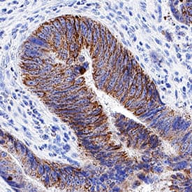
![Immunohistochemistry-Paraffin: SERCA3 ATPase Antibody (2H3) [H00000489-M01] - Analysis of monoclonal antibody to ATP2A3 on formalin-fixed paraffin-embedded human tonsil. Antibody concentration 3 ug/ml.](https://images.novusbio.com/fullsize/SERCA3-ATPase-Antibody-2H3-Immunohistochemistry-Paraffin-H00000489-M01-img0007.jpg)
Mouse Monoclonal
Species Human
Applications WB, ELISA, IHC
| 3 Publications |
|
![Knockdown Validated: Calreticulin Antibody [NB600-101] - Western blot shows lysates of HeLa human cervical epithelial carcinoma parental cell line and Calreticulin knockout (KO) HeLa cell line. PVDF membrane was probed with 1:1500 of Rabbit Anti-Human Calreticulin Polyclonal Antibody (Catalog # NB600-101) followed by HRP-conjugated Anti-Rabbit IgG Secondary Antibody (Catalog #HAF008). Specific band was detected for Calreticulin at approximately 55 kDa (as indicated) in the parental HeLa cell line, but is not detectable in the knockout HeLa cell line. This experiment was conducted under reducing conditions.](https://images.novusbio.com/fullsize/Calreticulin-Antibody-Knockdown-Validated-NB600-101-img0011.jpg)
![Western Blot: Calreticulin Antibody [NB600-101] - Human kidney lysate.](https://images.novusbio.com/fullsize/Calreticulin-Antibody-Western-Blot-NB600-101-img0006.jpg)
Rabbit Polyclonal
Species Human, Mouse, Rat
Applications WB, Simple Western, DB
| 28 Publications |
|
![Western Blot: ATP2C1 Antibody [NBP1-76566] - mouse brain tissue lysate with ATP2C1 antibody at 1 ug/mL in (A) the absence and (B) the presence of blocking peptide.](https://images.novusbio.com/fullsize/ATP2C1-Antibody-Western-Blot-NBP1-76566-img0001.jpg)
![Immunocytochemistry/Immunofluorescence: ATP2C1 Antibody [NBP1-76566] - Immunofluorescence of ATP2C1 in mouse brain tissue with ATP2C1 antibody at 20 ug/mL.](https://images.novusbio.com/fullsize/ATP2C1-Antibody-Immunocytochemistry-Immunofluorescence-NBP1-76566-img0002.jpg)
Rabbit Polyclonal
Species Human, Mouse
Applications WB, ELISA, ICC/IF
|
|
![Western Blot: SERCA1 ATPase Antibody (1B11) [H00000487-M01] - Analysis of ATP2A1 expression in transfected 293T cell line by ATP2A1 monoclonal antibody (M01), clone 1B11.Lane 1: ATP2A1 transfected lysate(109.3 KDa).Lane 2: Non-transfected lysate.](https://images.novusbio.com/fullsize/SERCA1-ATPase-Antibody-1B11-Western-Blot-H00000487-M01-img0011.jpg)
![Immunocytochemistry/Immunofluorescence: SERCA1 ATPase Antibody (1B11) [H00000487-M01] - Analysis of monoclonal antibody to ATP2A1 on A-431 cell. Antibody concentration 10 ug/ml.](https://images.novusbio.com/fullsize/SERCA1-ATPase-Antibody-1B11-Immunocytochemistry-Immunofluorescence-H00000487-M01-img0006.jpg)
Mouse Monoclonal
Species Human
Applications WB, ELISA, ICC/IF
|
|
![Western Blot: Involucrin Antibody (SY5) [NB100-2727] - Involucrin from primary human epidermal keratinocyte lysates grown in low calcium (0.07 mM) for 5 days or differentiated with high calcium (1.5 mM) for 1-5 days. Block in 5% nonfat dry milk in TBST incubated at RT for 1 hr, antibody 1:500 in TBST, overnight at 4C. Image from verified customer review.](https://images.novusbio.com/fullsize/Involucrin-Antibody-SY5-Western-Blot-NB100-2727-img0003.jpg)
![Immunohistochemistry-Paraffin: Involucrin Antibody (SY5) [NB100-2727] - Staining of human Tonsil with Involucrin Monoclonal Antibody (SY5).](https://images.novusbio.com/fullsize/Involucrin-Antibody-SY5-Immunohistochemistry-Paraffin-NB100-2727-img0002.jpg)
Mouse Monoclonal
Species Human, Porcine, Canine
Applications WB, Flow, ICC/IF
| 1 Review 2 Publications |
|
![Western Blot: Plasminogen Antibody [NB600-930] - Lane 1: Plasminogen. Lane 2: None. Load: 50 ng per lane. Primary antibody: Plasminogen primary antibody at 1:1,000 overnight at 4C. Secondary antibody: Peroxidase goat secondary antibody at 1:40,000 for 60 min at RT. Blocking: incubated with blocking buffer for 30 min at RT. Predicted/Observed size: 91 kDa, 91 kDa for Plasminogen. Other band(s): None.](https://images.novusbio.com/fullsize/Plasminogen-Antibody-Western-Blot-NB600-930-img0006.jpg)
![Western Blot: Plasminogen Antibody [NB600-930] - Detection of Plasminogen under reducing (R) and non-reducing (NR) conditions. Reduced samples of purified target proteins contained 4% BME and were boiled for 5 minutes. Samples of 1ug of protein per lane were run by SDS-PAGE. Protein was transferred to nitrocellulose and probed with 1:3000 dilution of primary antibody. Detection shown was using Dylight 649 conjugated Donkey anti goat 1 hr RT.](https://images.novusbio.com/fullsize/Plasminogen-Antibody-Western-Blot-NB600-930-img0005.jpg)
Goat Polyclonal
Species Human, Rat
Applications WB, ELISA
| 2 Reviews 6 Publications |
|


Mouse Monoclonal
Species Human, Mouse, Rat
Applications WB, Flow, ICC/IF
| 1 Review 59 Publications |
|


Goat Polyclonal
Species Human, Mouse
Applications WB, Simple Western, Flow
| 136 Publications |
|

Mouse Monoclonal
Species Human
Applications WB, IP
|
|

Goat Polyclonal
Species Human
Applications WB, IHC
| 4 Publications |
|
![Immunohistochemistry-Paraffin: Cytokeratin 17 Antibody [NBP2-38536] - Analysis in human urinary bladder and liver tissues using NBP2-38536 antibody. Corresponding Cytokeratin 17 RNA-seq data are presented for the same tissues.](https://images.novusbio.com/fullsize/Cytokeratin-17-Antibody-Immunohistochemistry-Paraffin-NBP2-38536-img0012.jpg)
![Western Blot: Cytokeratin 17 Antibody [NBP2-38536] - Analysis in human cell lines HeLa and PC-3. Corresponding RNA-seq data are presented for the same cell lines. Loading control: Anti-HSP90B1.](https://images.novusbio.com/fullsize/Cytokeratin-17-Antibody-Western-Blot-NBP2-38536-img0011.jpg)
Rabbit Polyclonal
Species Human
Applications WB, ICC/IF, IHC
| 1 Review |
|
![Immunohistochemistry-Paraffin: Desmoplakin Antibody [NBP2-48836] - Staining in human skin and skeletal muscle tissues using anti-DSP antibody. Corresponding DSP RNA-seq data are presented for the same tissues.](https://images.novusbio.com/fullsize/Desmoplakin-Antibody-Immunohistochemistry-Paraffin-NBP2-48836-img0005.jpg)
![Immunohistochemistry-Paraffin: Desmoplakin Antibody [NBP2-48836] - Staining of human skeletal muscle shows low expression as expected.](https://images.novusbio.com/fullsize/Desmoplakin-Antibody-Immunohistochemistry-Paraffin-NBP2-48836-img0003.jpg)
Rabbit Polyclonal
Species Human, Canine
Applications ICC/IF, IHC, IHC-P
| 2 Publications |
|
![Western Blot: Cytokeratin 10 Antibody (3C2F5) [NBP2-61736] - Analysis using KRT10 mAb against HEK293 (1) and KRT10 (AA: 345-454)-hIgGFc transfected HEK293 (2) cell lysate.](https://images.novusbio.com/fullsize/Cytokeratin-10-Antibody-3C2F5-Western-Blot-NBP2-61736-img0006.jpg)
![Immunohistochemistry-Paraffin: Cytokeratin 10 Antibody (3C2F5) [NBP2-61736] - Analysis of esophageal cancer tissues using KRT10 mouse mAb with DAB staining.](https://images.novusbio.com/fullsize/Cytokeratin-10-Antibody-3C2F5-Immunohistochemistry-NBP2-61736-img0004.jpg)
Mouse Monoclonal
Species Human, Mouse, Rat
Applications WB, ELISA, Flow
| 3 Publications |
|
![Western Blot: Cytokeratin 16 Antibody (SC52-09) [NBP2-67559] - Analysis of Cytokeratin 16 on human skin lysates using anti-Cytokeratin 16 antibody at 1/1,000 dilution.](https://images.novusbio.com/fullsize/Cytokeratin-16-Antibody-SC52-09-Western-Blot-NBP2-67559-img0008.jpg)
![Immunocytochemistry/Immunofluorescence: Cytokeratin 16 Antibody (SC52-09) [NBP2-67559] - Staining Cytokeratin 16 in HepG2 cells (green). The nuclear counter stain is DAPI (blue). Cells were fixed in paraformaldehyde, permeabilised with 0.25% Triton X100/PBS.](https://images.novusbio.com/fullsize/Cytokeratin-16-Antibody-SC52-09-Immunocytochemistry-Immunofluorescence-NBP2-67559-img0004.jpg)
Rabbit Monoclonal
Species Human
Applications WB, Flow, ICC/IF
|
|
![Western Blot: AKR1C2 Antibody (CPTC-AKR1C2-1) [NBP2-79775] - Western Blot Analysis of Human HeLa, K-562 and A431 cell lysates using AKR1C2 Antibody (CPTC-AKR1C2-1).](https://images.novusbio.com/fullsize/AKR1C2-Antibody-CPTC-AKR1C2-1-Western-Blot-NBP2-79775-img0004.jpg)
![Immunohistochemistry-Paraffin: AKR1C2 Antibody (CPTC-AKR1C2-1) [NBP2-79775] - Formalin-fixed, paraffin-embedded human Prostate Carcinoma stained with AKR1C2 Antibody (CPTC-AKR1C2-1).](https://images.novusbio.com/fullsize/AKR1C2-Antibody-CPTC-AKR1C2-1-Immunohistochemistry-Paraffin-NBP2-79775-img0002.jpg)
Mouse Monoclonal
Species Human
Applications WB, IHC, IHC-P
|
|
![Western Blot: SAT1 Antibody [NB110-41622] - Detection of SAT1 in human SAT1 transfected lysate.](https://images.novusbio.com/fullsize/SAT1-Antibody-Western-Blot-NB110-41622-img0004.jpg)
![Immunohistochemistry: SAT1 Antibody [NB110-41622] - IHC analysis of SAT1 in mouse seminal vesical (left) and prostate (right) usind DAB with hematoxylin counterstain,](https://images.novusbio.com/fullsize/SAT1-Antibody-Immunohistochemistry-NB110-41622-img0006.jpg)
Rabbit Polyclonal
Species Human, Mouse, Rat
Applications WB, IHC, IHC-P
| 5 Publications |
|
![Western Blot: DSPP Antibody [NBP2-92546] - Analysis of extracts of various cell lines, using DSPP at 1:1000 dilution.Secondary antibody: HRP Goat Anti-Rabbit IgG (H+L) at 1:10000 dilution.Lysates/proteins: 25ug per lane.Blocking buffer: 3% nonfat dry milk in TBST.Detection: ECL Basic Kit .Exposure time: 30s.](https://images.novusbio.com/fullsize/DSPP-Antibody-Western-Blot-NBP2-92546-img0004.jpg)
Rabbit Polyclonal
Species Human, Mouse, Rat
Applications WB
| 3 Publications |
|
![Immunocytochemistry/Immunofluorescence: SERCA2 ATPase Antibody (2A7-A1) [NB300-581] - HeLa cells. Primary antibody at 1:100, secondary antibody: goat-anti mouse IgG AlexaFluor 568 at 1:100. ICC/IF image submitted by a verified customer review.](https://images.novusbio.com/fullsize/SERCA2-ATPase-Antibody-2A7-A1-Immunocytochemistry-Immunofluorescence-NB300-581-img0010.jpg)
![Immunohistochemistry-Paraffin: SERCA2 ATPase Antibody (2A7-A1) [NB300-581] - Both normal and cancer biopsies of deparaffinized Human skeletal muscle tissues.](https://images.novusbio.com/fullsize/SERCA2-ATPase-Antibody-2A7-A1-Immunohistochemistry-Paraffin-NB300-581-img0008.jpg)
Mouse Monoclonal
Species Human, Mouse, Rat
Applications WB, Flow, ICC/IF
| 2 Reviews 16 Publications |
|
