| Submit your blog on Epispadias to be featured! |
| Submit your event on Epispadias to be featured! |
![Western Blot: UVRAG Antibody [NBP1-18885] - Beclin 1 is acetylated at lysines 430 and 437. TSA and NAM increase the binding of Beclin 1 to Rubicon. Immunoprecipitation of indicated Beclin 1-binding partners with ectopically expressed Flag-Beclin 1 in HEK293T cells treated with TSA and NAM. Image collected and cropped by CiteAb from the following publication (https://www.nature.com/articles/ncomms8215), licensed under a CC-BY license.](https://images.novusbio.com/fullsize/UVRAG-Antibody-Western-Blot-NBP1-18885-img0005.jpg)
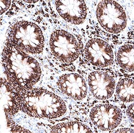
![Immunohistochemistry-Paraffin: UVRAG Antibody [NBP1-18885] - Section of human colon carcinoma. Antibody: Affinity purified rabbit anti- UVRAG used at a dilution of 1:200 (1ug/ml). Detection: DAB](https://images.novusbio.com/fullsize/UVRAG-Antibody-Immunohistochemistry-Paraffin-NBP1-18885-img0004.jpg)
Rabbit Polyclonal
Species Human
Applications WB, ICC/IF, IHC
| 3 Publications |
|
Mouse Monoclonal
Species Human
Applications WB, Simple Western, IHC
|
|
![Immunohistochemistry: Neuropeptide Y Antibody [NBP1-46535] - Low magnification images showing the double staining pattern of NHE1 with neuropeptide NPY. Scale bar: 100um. Insets: High magnification images showing individual cells with double staining. Scale bar: 10um. Asterisks mark Hoechst-stained nuclei. Image collected and cropped by CiteAb from the following publication (nature.com/articles/s41598-019-42872-w), licensed under a CC-BY license.](https://images.novusbio.com/fullsize/Neuropeptide-Y-Antibody-Immunohistochemistry-NBP1-46535-img0001.jpg)
![Immunocytochemistry/ Immunofluorescence: Neuropeptide Y Antibody [NBP1-46535] - NHE1 distribution (A) & colocalisation with markers for specific cell types (B–D) & major inputs (E, F, G). (A) NHE1 immunoreactivity is distributed throughout the rostrocaudal axis of the SCN (encircled by the dotted lines). Scale bar: 200 µm. OC: optic chiasm. 3 V: third ventricle. (B–G1) Low magnification images showing the double staining pattern of NHE1 with neuropeptides NP2 (B), GRP (C), & VIP (D) as well as markers for afferent inputs vGluT2 (E), NPY (F), & SERT (G1). Scale bar: 100 µm. Insets: High magnification images showing individual cells with double staining. Scale bar: 10 µm. Asterisks mark Hoechst-stained nuclei. (G2) High magnification image showing high degree of colocalisation (yellow) between NHE1 (green) & SERT (red). Scale bar: 10 µm. Image collected & cropped by CiteAb from the following publication (https://pubmed.ncbi.nlm.nih.gov/31015514), licensed under a CC-BY license. Not internally tested by Novus Biologicals.](https://images.novusbio.com/fullsize/nbp1-46535_goat-polyclonal-neuropeptide-y-antibody-310202416161848.jpg)
Goat Polyclonal
Species Human, Mouse, Rat
Applications WB, ICC/IF, IHC
| 20 Publications |
|
![Western Blot: Pancreatic Amylase Alpha Antibody (6D4) [NBP1-47659] - Pancreatic Amylase Alpha Antibody (6D4) HEK293T cells were transfected with the pCMV6-ENTRY control (Left lane) or pCMV6-ENTRY Pancreatic Amylase(Right lane) cDNA for 48 hrs and lysed. Equivalent amounts of cell lysates (5 ug per lane) were separated by SDS-PAGE and immunoblotted with anti-Pancreatic Amylase.](https://images.novusbio.com/fullsize/Pancreatic-Amylase-Alpha-Antibody-6D4-Western-Blot-NBP1-47659-img0022.jpg)
![Immunohistochemistry-Paraffin: Pancreatic Amylase Alpha Antibody (6D4) [NBP1-47659] - Pancreatic Amylase Alpha Antibody (6D4) Staining of paraffin-embedded ovary using anti-Pancreatic Amylase mouse monoclonal antibody.](https://images.novusbio.com/fullsize/Pancreatic-Amylase-Alpha-Antibody-6D4-Immunohistochemistry-Paraffin-NBP1-47659-img0021.jpg)
Mouse Monoclonal
Species Human
Applications WB, IHC, IHC-P
| 1 Review |
|
![Western Blot: SF-1/NR5A1/Steroidogenic Factor 1 Antibody [NBP1-52823] - Titration: 0.2-1 ug/ml, Positive Control: THP-1 cell lysate.](https://images.novusbio.com/fullsize/SF-1-NR5A1-Steroidogenic-Factor-1-Antibody-Western-Blot-NBP1-52823-img0007.jpg)
![Immunohistochemistry-Paraffin: SF-1/NR5A1/Steroidogenic Factor 1 Antibody [NBP1-52823] - Human adrenal tissue at an antibody concentration of 4-8ug/ml.](https://images.novusbio.com/fullsize/SF-1-NR5A1-Steroidogenic-Factor-1-Antibody-Immunohistochemistry-Paraffin-NBP1-52823-img0006.jpg)
Rabbit Polyclonal
Species Human, Mouse
Applications WB, Simple Western, ICC/IF
| 4 Publications |
|
![Western Blot: PP14/Glycodelin Antibody [NBP1-89781] - Analysis in human placenta tissue.](https://images.novusbio.com/fullsize/PP14-Glycodelin-Antibody-Western-Blot-NBP1-89781-img0014.jpg)
![Immunohistochemistry-Paraffin: PP14/Glycodelin Antibody [NBP1-89781] - Staining of human testis shows no positivity in cells in seminiferous ducts as expected.](https://images.novusbio.com/fullsize/PP14-Glycodelin-Antibody-Immunohistochemistry-Paraffin-NBP1-89781-img0019.jpg)
Rabbit Polyclonal
Species Human
Applications WB, IHC, IHC-P
|
|
![Western Blot: Androgen R/NR3C4 [p Ser213, p Ser210] Antibody (156C135.2) [NB100-56603] - Analysis using Azide Free version of NB100-56603. LNCaP cells (passage number 38) were serum-starved for 2 days. After serum starvation, cells were (A) left untreated, (B) treated with 100 ng/ml IGF-1 for 4h, or (C) incubated with 20 um LY294002 for 30 mi](https://images.novusbio.com/fullsize/Androgen-R-NR3C4-p-Ser213-p-Ser210-Antibody-156C135-2-Western-Blot-NB100-56603-img0004.jpg)
Mouse Monoclonal
Species Human, Mouse, Rat
Applications WB, IHC, IHC-P
| 14 Publications |
|
![Immunohistochemistry-Paraffin: Capicua Antibody [NBP2-33420] - Staining in human testis and liver tissues . Corresponding CIC RNA-seq data are presented for the same tissues.](https://images.novusbio.com/fullsize/Capicua-Antibody-Immunohistochemistry-Paraffin-NBP2-33420-img0011.jpg)
![Western Blot: Capicua Antibody [NBP2-33420] - Analysis in A-549 cells transfected with control siRNA, target specific siRNA probe #1 and #2, using anti-CIC antibody. Remaining relative intensity is presented. Loading control: anti-GAPDH.](https://images.novusbio.com/fullsize/Capicua-Antibody-Western-Blot-NBP2-33420-img0013.jpg)
Rabbit Polyclonal
Species Human
Applications WB, ICC/IF, IHC
|
|


Goat Polyclonal
Species Human
Applications WB, IHC, ICC
| 1 Review 24 Publications |
|
![Western Blot: PLOD1 Antibody [NBP2-38770] - Analysis in human cell line U-251 MG and human cell line RT-4.](https://images.novusbio.com/fullsize/PLOD1-Antibody-Western-Blot-NBP2-38770-img0006.jpg)
![Western Blot: PLOD1 Antibody [NBP2-38770] - SC65 directly interacts with lysyl-hydroxylase 1 (LH1). Western blot of primary calvarial osteoblast and skin fibroblast lysates from WT and Sc65KO 3 day-old mice (N = 2) showing significantly decreased levels of LH1 protein in Sc65KO samples. Densitometric quantification of LH1 protein normalized to beta-actin from the western blot shown above (*p<0.05; error bars represent SD). All experiments were performed at least 3 times. Image collected and cropped by CiteAb from the following publication (https://dx.plos.org/10.1371/journal.pgen.1006002), licensed under a CC-BY license.](https://images.novusbio.com/fullsize/PLOD1-Antibody-Western-Blot-NBP2-38770-img0005.jpg)
Rabbit Polyclonal
Species Human, Mouse
Applications WB, IHC-P
| 3 Publications |
|


Species Human
Applications BA
| 103 Publications |
|


Species Human
Applications BA
| 848 Publications |
|
![Immunohistochemistry-Paraffin: Desmoplakin Antibody [NBP2-48836] - Staining in human skin and skeletal muscle tissues using anti-DSP antibody. Corresponding DSP RNA-seq data are presented for the same tissues.](https://images.novusbio.com/fullsize/Desmoplakin-Antibody-Immunohistochemistry-Paraffin-NBP2-48836-img0005.jpg)
![Immunohistochemistry-Paraffin: Desmoplakin Antibody [NBP2-48836] - Staining of human skeletal muscle shows low expression as expected.](https://images.novusbio.com/fullsize/Desmoplakin-Antibody-Immunohistochemistry-Paraffin-NBP2-48836-img0003.jpg)
Rabbit Polyclonal
Species Human, Canine
Applications ICC/IF, IHC, IHC-P
| 2 Publications |
|
![Western Blot: RPE65 Antibody (401.8B11.3D9) - BSA Free [NB100-355] - Subcellular localization of BEST1 and surface Ca2+-dependent Cl- current in patient-derived iPSC-RPEs. Western blots show similar BEST1 expression levels in WT and patient-derived iPSC-RPEs. Each sample was from one cell lysis (BEST1 and beta-actin, RPE65 and CRALBP were on two gels, respectively). Image collected and cropped by CiteAb from the following publication (https://elifesciences.org/articles/29914), licensed under a CC-BY license.](https://images.novusbio.com/fullsize/RPE65-Antibody-401-8B11-3D9-BSA-Free-Western-Blot-NB100-355-img0013.jpg)
![Immunocytochemistry/Immunofluorescence: RPE65 Antibody (401.8B11.3D9) - BSA Free [NB100-355] - Expression of eye-specific markers in the induced eye-like structures induced from lignin-added ES cells. (a) Higher-magnification image of the RPE like structure induced from ESCs after 12-day culture. (b) Immunostaining of eye- like structures. Eye- like structures induced from ESCs after 12-day culture were stained with antibodies against RPE65 (red) and nuclei were stained with DAPI solution (blue). Scale bar: a = 50 um, b,d = 200 um, c,e = 100 um. PLoS One. 2013 Jun 21;8(6):e66376. doi: 10.1371/journal.pone.0066376.](https://images.novusbio.com/fullsize/RPE65-Antibody-401-8B11-3D9-BSA-Free-Immunocytochemistry-Immunofluorescence-NB100-355-img0014.jpg)
Mouse Monoclonal
Species Human, Mouse, Rat
Applications WB, Simple Western, Flow
| 7 Reviews 98 Publications |
|
Mouse Monoclonal
Species Human, Rat
Applications WB, ICC/IF, IHC
| 1 Review 11 Publications |
|
![Western Blot: Gastrin-releasing Peptide/Bombesin/Neuromedin C Antibody [NBP3-03282] - Analysis of extracts of various cell lines, using Gastrin-releasing Peptide/Bombesin/Neuromedin C antibody (NBP3-03282) at 1:1000 dilution. Secondary antibody: HRP Goat Anti-Rabbit IgG (H+L) at 1:10000 dilution. Lysates/proteins: 25ug per lane. Blocking buffer: 3% nonfat dry milk in TBST. Detection: ECL Basic Kit. Exposure time: 180s.](https://images.novusbio.com/fullsize/Gastrin-releasing-Peptide-Bombesin-Neuromedin-C-Antibody-Western-Blot-NBP3-03282-img0004.jpg)
![Immunocytochemistry/Immunofluorescence: Gastrin-releasing Peptide/Bombesin/Neuromedin C Antibody [NBP3-03282] - Immunofluorescence analysis of HeLa cells using Gastrin-releasing Peptide/Bombesin/Neuromedin C antibody (NBP3-03282) at dilution of 1:100. Blue: DAPI for nuclear staining.](https://images.novusbio.com/fullsize/Gastrin-releasing-Peptide-Bombesin-Neuromedin-C-Antibody-Immunocytochemistry-Immunofluorescence-NBP3-03282-img0001.jpg)
Rabbit Polyclonal
Species Human, Mouse, Rat
Applications WB, ICC/IF, IHC
|
|
![Western Blot: alpha Desmuslin Antibody [NBP3-13366] - Mouse tissue extract (50 ug) was separated by 5% SDS-PAGE, and the membrane was blotted with alpha Desmuslin antibody (NBP3-13366) diluted at 1:1000. The HRP-conjugated anti-rabbit IgG antibody (NBP2-19301) was used to detect the primary antibody.](https://images.novusbio.com/fullsize/alpha-Desmuslin-Antibody-Western-Blot-NBP3-13366-img0002.jpg)
![Immunocytochemistry/Immunofluorescence: alpha Desmuslin Antibody [NBP3-13366] - alpha Desmuslin antibody detects alpha Desmuslin protein by immunofluorescent analysis. Sample: DIV10 rat E18 primary cortical neuron and glia cells were fixed in 4% paraformaldehyde at RT for 15 min. Green: alpha Desmuslin stained by alpha Desmuslin antibody (NBP3-13366) diluted at 1:500. Blue: Fluoroshield with DAPI.](https://images.novusbio.com/fullsize/alpha-Desmuslin-Antibody-Immunocytochemistry-Immunofluorescence-NBP3-13366-img0001.jpg)
Rabbit Polyclonal
Species Mouse, Rat
Applications WB, ICC/IF, IHC
|
|
