 |
By Jamshed Arslan, Pharm. D., PhD.
Novel coronavirus or SARS-CoV-2 has gripped the entire world. Infected individuals keep shedding the virus in respiratory droplets even before showing symptoms because of high viral load in their upper respiratory tract. The virus is also detectable in blood and feces of the infected people. COVID-19 symptoms mainly include fever, fatigue, dry cough and difficulty breathing, which may develop into severe pneumonia. To understand this disease, it is crucial to recapitulate key aspects of COVID-19 respiratory pathology in evolutionarily related animals where repeated sampling could be feasible.
A team at the National Institutes of Health (NIH) , USA, that had previously presented rhesus macaques as a model for a related coronavirus MERS-CoV, has now generated a rhesus macaque model to represent SARS-CoV-2 infection in humans. Their model successfully reflects the underlying clinical and respiratory pathology of moderate COVID-19. To reliably detect SARS-CoV-2 antigen in all their samples, the NIH team used anti-SARS-CoV nucleocapsid antibody to evaluate through immunohistochemistry the presence of the virus which was found in type 1 and 2 pneumocytes, alveolar macrophages, mediastinal lymph node, and macrophages and lymphocytes in the lamina propria of the cecum.

|
SARS-CoV-2 antigen was detected by immunohistochemistry using a SARS nucleocapsid rabbit polyclonal antibody [NB100-56576] at a dilution of 1:250. Positive detection of SARS nucleocapsid antigen is shown in (g) type I pneumocytes, (j) type I and type II pneumocytes and alveolar macrophages, (k) mediastinal lymph node, and (l) macrophages and lymphocytes in the lamina propria of the cecum. Images obtained from Respiratory disease and virus shedding in rhesus macaques inoculated with SARS-CoV-2 Figure 4, modified diagram shows only a section of Figure 4. |
SARS-CoV Nucleocapsid Protein Antibodies at Novus Biologicals |
|

|
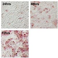
SARS Nucleocapsid Protein Antibody [NB100-56576] - Characteristics of 2B4 cells clonally derived from human bronchial epithelial Calu-3 cells. Finally, 2B4 cells (passage 6) were infected with SARS-CoV (MOI = 0.1) for 24, 48, and 72 hrs before being fixed with 4% paraformaldehyde for monitoring the morphological changes of infected cells, as visualized by the expression of SARS-CoV NP protein (red) by using the standard IHC (F). Image collected and cropped by CiteAb from PLOS licensed under a CC-BY license. |
View SARS Nucleocapsid Antibodies
To represent COVID-19 in our evolutionary cousins, the researchers infected 8 monkeys with SARS-CoV-2 via nose, trachea, eyes and/or mouth, and studied them for 3 weeks. Symptoms like fever, irregular breathing, reduced appetite, pale appearance and dehydration developed. Radiographs showed pulmonary infiltrates that resolved within 2 weeks. The team euthanized 4 animals each on 3 and 21 days post-infection (dpi) and found interstitial pneumonia and multi-focal lesions. All respiratory tissues showed viral mRNA, indicating active virus replication, on 3 dpi, but not on 21 dpi. Overall, the presence of pulmonary infiltrates, virus replication pattern and histological lesions resembled human COVID-19 progression.
To estimate viral loads, swabs were taken from nose, throat, rectum and urogenital area. Nasal virus shedding was the highest among all. Bronchoalveolar fluids showed high viral load whereas viral RNA could not be detected in blood, urine or central nervous system. Throat swabs showed high viral load after the infection, but the virus levels were inconsistent. One animal kept shedding viral RNA in rectal swabs even after 2 weeks.
Overall, the pattern of virus shedding from respiratory and intestinal tracts was similar to that seen in humans. ELISA on 10 dpi revealed that all four animals were seropositive for SARS-CoV-2 spike with IgG titers closer to those seen in the patients. However, the scientists failed to observe a reduction in viral load immediately after seroconversion as occurs in COVID-19 humans.
The importance of representing COVID-19 in a primate cannot be overstated. A monkey model allows frequent tissue collection for a detailed study of COVID-19 pathology. Likewise, this research will open doors to vaccines and anti-viral therapies against SARS-CoV-2. In fact, the same NIH team stated that "we have … moved forward to test antiviral treatments and vaccines in this model."
Explore Bio-Techne’s Resources for SARS-CoV-2 Research »
 Jamshed Arslan, Pharm D, PhD
Dr Arslan is an Assistant Professor at Barrett Hodgson University, Pakistan,
where he uses various pedagogical methods to teach Pharm D students.
Research in focus
Munster VJ, Feldmann F, Williamson BN , Doremalen N, Pérez-Pérez L, Schulz J, Meade-White K,… de Wit E. (2020). Respiratory Disease and Virus Shedding in Rhesus Macaques Inoculated with SARS-CoV-2. BioRxiv [Pre-print]. https://doi.org/10.1101/2020.03.21.001628