
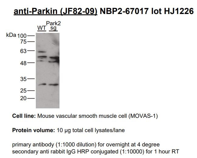
![Western Blot: Parkin Antibody (JF82-09) [NBP2-67017] - Analysis of Parkin on 293T cell lysates. Proteins were transferred to a PVDF membrane and blocked with 5% BSA in PBS for 1 hour at room temperature. The primary antibody (1/500) was used in 5% BSA at room temperature for 2 hours. Goat Anti-Rabbit IgG - HRP Secondary Antibody at 1:5,000 dilution was used for 1 hour at room temperature.](http://images.novusbio.com/fullsize/Parkin-Antibody-JF82-09-Western-Blot-NBP2-67017-img0007.jpg "Western Blot: Parkin Antibody (JF82-09) [NBP2-67017] - Analysis of Parkin on 293T cell lysates. Proteins were transferred to a PVDF membrane and blocked with 5% BSA in PBS for 1 hour at room temperature. The primary antibody (1/500) was used in 5% BSA at room temperature for 2 hours. Goat Anti-Rabbit IgG - HRP Secondary Antibody at 1:5,000 dilution was used for 1 hour at room temperature.")
| Reactivity | Hu, Mu, RtSpecies Glossary |
| Applications | WB, Flow, ICC/IF, IHC, IP |
| Clone | JF82-09 |
| Clonality | Monoclonal |
| Host | Rabbit |
| Conjugate | Unconjugated |
| Concentration | 1 mg/ml |
| Additional Information | Recombinant Monoclonal Antibody. |
| Immunogen | Synthetic peptide within N-terminal human Parkin. (SwissProt: O60260 Human; SwissProt: Q9WVS6 Mouse; SwissProt: Q9JK66 Rat) |
| Localization | Mitochondrion, mitochondrion outer membrane, endoplasmic reticulum, cytosol, Nucleus, neuron projection, postsynaptic density, presynapse. |
| Isotype | IgG |
| Clonality | Monoclonal |
| Host | Rabbit |
| Gene | PRKN |
| Purity | Protein A purified |
| Innovator's Reward | Test in a species/application not listed above to receive a full credit towards a future purchase. |
| Dilutions |
|
|
| Theoretical MW | 52 kDa. Disclaimer note: The observed molecular weight of the protein may vary from the listed predicted molecular weight due to post translational modifications, post translation cleavages, relative charges, and other experimental factors. |
|
| Reviewed Applications |
|
|
| Publications |
|
| Storage | Store at 4C short term. Aliquot and store at -20C long term. Avoid freeze-thaw cycles. |
| Buffer | TBS (pH7.4), 0.05% BSA, 40% Glycerol |
| Preservative | 0.05% Sodium Azide |
| Concentration | 1 mg/ml |
| Purity | Protein A purified |


")
| Images | Ratings | Applications | Species | Date | Details | ||||||||
|---|---|---|---|---|---|---|---|---|---|---|---|---|---|
|
reviewed by:
Verified Customer |
ICC | Human | 02/10/2022 |
Summary
|
|||||||||
Enlarge |
reviewed by:
Verified Customer |
ICC | Human | 02/21/2019 |
Summary
|
||||||||
Enlarge |
reviewed by:
Verified Customer |
WB | Mouse | 12/21/2018 |
Summary
|
Secondary Antibodies |
Isotype Controls |
Research Areas for Parkin Antibody (NBP2-67017)Find related products by research area.
|
|
Understanding Mitophagy Mechanisms: Canonical PINK1/Parkin, LC3-Dependent Piecemeal, and LC3-Independent Mitochondrial Derived Vesicles By Christina Towers, PhD What is Mitophagy?The selective degradation of mitochondria via double membrane autophagosome vesicles is called mitophagy. Damaged mitochondria can generate harmful amounts of reactive ox... Read full blog post. |
|
New Players in the Mitophagy Game By Christina Towers, PhD Mitochondrial turn over via the lysosome, otherwise known as mitophagy, involves engulfment of mitochondria into double membrane autophagosomes and subsequent fusion with lysosomes. Much is al... Read full blog post. |
|
Optogenetic Control of Mitophagy: AMBRA1 based mitophagy switch By Christina Towers, PhD Mitophagy in the BrainSelective autophagic degradation of damaged mitochondria, known as mitophagy, has been described as a cyto-protective process. Accordingly, defects in mitophagy h... Read full blog post. |
|
There's an autophagy for that! By Christina Towers, PhDA critical mechanism that cells use to generate nutrients and fuel metabolism is through a process called autophagy. This process is complex and involves over 20 different proteins, most of which are highly conserved acro... Read full blog post. |
|
The role of Parkin and autophagy in retinal pigment epithelial cell (RPE) degradation The root of Parkinson’s disease (PD) points to a poorly regulated electron transport chain leading to mitochondrial damage, where many proteins need to work cohesively to ensure proper function. The two key players of this pathway are PINK1, ... Read full blog post. |
|
Parkin - Role in Mitochondrial Quality Control and Parkinson's Disease Parkin/PARK2 is a cytosolic enzyme which gets recruited to cellular mitochondria damaged through depolarization, ROS or unfolded proteins accumulation, and exert protective effects by inducing mitophagy (mitochondrial autophagy). Parkin induces mit... Read full blog post. |
|
PINK1 - performing mitochondrial quality control and protecting against Parkinson’s disease PTEN-induced putative kinase 1 (PINK1) is a serine/threonine kinase with important functions in mitochondrial quality control. Together with the Parkin protein, PINK1 is able to regulate the selective degradation of damaged mitochondria through aut... Read full blog post. |
|
PINK1: All work and no fun The protein PINK1 is a mitochondrial-located serine/threonine kinase (PTK) that maintains organelle function and integrity. It not only protects organelles from cellular stress, but it also uses the selective auto-phagocytosis process for cleaning and... Read full blog post. |
|
PINK1: Promoting Organelle Stability and Preventing Parkinson's disease PINK1 is a protein serine/threonine kinase (PTK) that protects the organelles from cellular stress and controls selective autophagy to clear damage. Exner, et al. were among the first to report that PINK1 deficiency in humans was linked to autosomal r... Read full blog post. |
|
Novus Antibodies Highlighted in Parkinson's Disease Research Identified almost two centuries ago, Parkinson's disease is a neurodegenerative disorder that afflicts an estimated 4-6 million worldwide (www.parkinsons.org). The prevalence of Parkinson's disease is expected to grow considerably as the average age o... Read full blog post. |

The concentration calculator allows you to quickly calculate the volume, mass or concentration of your vial. Simply enter your mass, volume, or concentration values for your reagent and the calculator will determine the rest.
5 | |
4 | |
3 | |
2 | |
1 |
| Verified Customer 02/10/2022 |
||
| Application: | ICC | |
| Species: | Human |
| Verified Customer 02/21/2019 |
||
| Application: | ICC | |
| Species: | Human |
| Verified Customer 12/21/2018 |
||
| Application: | WB | |
| Species: | Mouse |
| Gene Symbol | PRKN |


![Immunohistochemistry-Paraffin: Parkin Antibody (JF82-09) [NBP2-67017] - Analysis of paraffin-embedded rat brain tissue using anti-Parkin antibody. Counter stained with hematoxylin.](http://images.novusbio.com/fullsize/Parkin-Antibody-JF82-09-Immunohistochemistry-Paraffin-NBP2-67017-img0004.jpg "Immunohistochemistry-Paraffin: Parkin Antibody (JF82-09) [NBP2-67017] - Analysis of paraffin-embedded rat brain tissue using anti-Parkin antibody. Counter stained with hematoxylin.")
![Flow Cytometry: Parkin Antibody (JF82-09) [NBP2-67017] - Analysis of SH-SY-5Y cells with Parkin antibody at 1/50 dilution (red) compared with an unlabelled control (cells without incubation with primary antibody; black). Alexa Fluor 488-conjugated goat anti rabbit IgG was used as the secondary antibody](http://images.novusbio.com/fullsize/Parkin-Antibody-JF82-09-Flow-Cytometry-NBP2-67017-img0001.jpg "Flow Cytometry: Parkin Antibody (JF82-09) [NBP2-67017] - Analysis of SH-SY-5Y cells with Parkin antibody at 1/50 dilution (red) compared with an unlabelled control (cells without incubation with primary antibody; black). Alexa Fluor 488-conjugated goat anti rabbit IgG was used as the secondary antibody")
![Immunocytochemistry/Immunofluorescence: Parkin Antibody (JF82-09) [NBP2-67017] - Staining Parkin in SH-SY-5Y cells (red). The nuclear counter stain is DAPI (blue). Cells were fixed in paraformaldehyde, permeabilised with 0.25% Triton X100/PBS.](http://images.novusbio.com/fullsize/Parkin-Antibody-JF82-09-Immunocytochemistry-Immunofluorescence-NBP2-67017-img0002.jpg "Immunocytochemistry/Immunofluorescence: Parkin Antibody (JF82-09) [NBP2-67017] - Staining Parkin in SH-SY-5Y cells (red). The nuclear counter stain is DAPI (blue). Cells were fixed in paraformaldehyde, permeabilised with 0.25% Triton X100/PBS.")
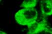
![Immunocytochemistry/Immunofluorescence: Parkin Antibody (JF82-09) [NBP2-67017] - Staining Parkin in PC-3M cells (red). The nuclear counter stain is DAPI (blue). Cells were fixed in paraformaldehyde, permeabilised with 0.25% Triton X100/PBS.](http://images.novusbio.com/fullsize/Parkin-Antibody-JF82-09-Immunocytochemistry-Immunofluorescence-NBP2-67017-img0005.jpg "Immunocytochemistry/Immunofluorescence: Parkin Antibody (JF82-09) [NBP2-67017] - Staining Parkin in PC-3M cells (red). The nuclear counter stain is DAPI (blue). Cells were fixed in paraformaldehyde, permeabilised with 0.25% Triton X100/PBS.")
![Immunocytochemistry/Immunofluorescence: Parkin Antibody (JF82-09) [NBP2-67017] - Staining Parkin in N2A cells (red). The nuclear counter stain is DAPI (blue). Cells were fixed in paraformaldehyde, permeabilised with 0.25% Triton X100/PBS.](http://images.novusbio.com/fullsize/Parkin-Antibody-JF82-09-Immunocytochemistry-Immunofluorescence-NBP2-67017-img0006.jpg "Immunocytochemistry/Immunofluorescence: Parkin Antibody (JF82-09) [NBP2-67017] - Staining Parkin in N2A cells (red). The nuclear counter stain is DAPI (blue). Cells were fixed in paraformaldehyde, permeabilised with 0.25% Triton X100/PBS.")
![Immunohistochemistry-Paraffin: Parkin Antibody (JF82-09) [NBP2-67017] - Analysis of paraffin-embedded mouse testis tissue using anti-Parkin antibody. Counter stained with hematoxylin.](http://images.novusbio.com/fullsize/Parkin-Antibody-JF82-09-Immunohistochemistry-Paraffin-NBP2-67017-img0003.jpg "Immunohistochemistry-Paraffin: Parkin Antibody (JF82-09) [NBP2-67017] - Analysis of paraffin-embedded mouse testis tissue using anti-Parkin antibody. Counter stained with hematoxylin.")
![Western Blot: Parkin Antibody (JF82-09) [NBP2-67017] -Analysis of Parkin on different lysates with Rabbit anti-Parkin antibody at 1/1,000 dilution.Lane 1: 293 cell lysateLane 2: Jurkat cell lysateLysates/proteins at 10 ug/Lane.Predicted band size: 52 kDaObserved band size: 52 kDaExposure time: 1 minutes;10% SDS-PAGE gel.Proteins were transferred to a PVDF membrane and blocked with 5% NFDM/TBST for 1 hour at room temperature. The primary antibody at 1/1,000 dilution was used in 5% NFDM/TBST at room temperature for 2 hours. Goat Anti-Rabbit IgG - HRP Secondary Antibody at 1:300,000 dilution was used for 1 hour at room temperature.](http://images.novusbio.com/fullsize/nbp2-67017_rabbit-monoclonal-parkin-antibody-jf82-09-western-blot-1962024104114..jpg "Western Blot: Parkin Antibody (JF82-09) [NBP2-67017] -Analysis of Parkin on different lysates with Rabbit anti-Parkin antibody at 1/1,000 dilution.Lane 1: 293 cell lysateLane 2: Jurkat cell lysateLysates/proteins at 10 ug/Lane.Predicted band size: 52 kDaObserved band size: 52 kDaExposure time: 1 minutes;10% SDS-PAGE gel.Proteins were transferred to a PVDF membrane and blocked with 5% NFDM/TBST for 1 hour at room temperature. The primary antibody at 1/1,000 dilution was used in 5% NFDM/TBST at room temperature for 2 hours. Goat Anti-Rabbit IgG - HRP Secondary Antibody at 1:300,000 dilution was used for 1 hour at room temperature.")


![Western Blot: Goat anti-Rabbit IgG (H+L) Secondary Antibody [HRP] [NB7160] - Western blot showing vemurafenib treatment in BRAFV600E CRC cells inhibits fission mediator DRP1 with no significant effect on fusion proteins (Mfn1 & 2) using MFN-1 antibody (NBP1-51841) and corresponding secondary antibody, goat anti-rabbit IgG-HRP (NB7160). Image collected and cropped by CiteAb from the following publication (https://pubmed.ncbi.nlm.nih.gov/33738242).](https://images.novusbio.com/images/Goat-anti-Rabbit-IgG-H+L-Secondary-Antibody-HRP-Western-Blot-NB7160-img0001.jpg "Western Blot: Goat anti-Rabbit IgG (H+L) Secondary Antibody [HRP] [NB7160] - Western blot showing vemurafenib treatment in BRAFV600E CRC cells inhibits fission mediator DRP1 with no significant effect on fusion proteins (Mfn1 & 2) using MFN-1 antibody (NBP1-51841) and corresponding secondary antibody, goat anti-rabbit IgG-HRP (NB7160). Image collected and cropped by CiteAb from the following publication (https://pubmed.ncbi.nlm.nih.gov/33738242).")
 using a 1:1000 dilution of HRP-conjugated Anti-Rabbit IgG Secondary Antibody (Catalog # HAF008). This experiment was conducted under reducing conditions and using Immunoblot Buffer Group 1.")
![Flow Cytometry: Rabbit IgG Isotype Control [NBP2-24891] - Intracellular FACS analysis of mouse TLR6 polyclonal antibody (red), rabbit isotype control (green), RAW cells alone (shaded). Two micrograms of antibodies were used. Goat anti-rabbit FITC (Novus, 20302) was used as secondary.](https://images.novusbio.com/images/Rabbit--Mouse-IgG-Isotype-Control-Flow-Cytometry-NBP2-24891-img0001.jpg "Flow Cytometry: Rabbit IgG Isotype Control [NBP2-24891] - Intracellular FACS analysis of mouse TLR6 polyclonal antibody (red), rabbit isotype control (green), RAW cells alone (shaded). Two micrograms of antibodies were used. Goat anti-rabbit FITC (Novus, 20302) was used as secondary.")


