and Hoechst (blue). IHC-P image submitted by a verified customer review.")
| Reactivity | Hu, Mu, Rt, Po, Bv, RbSpecies Glossary |
| Applications | WB, ELISA, ICC/IF, IHC, IP, MA, PAGE, KD |
| Clonality | Polyclonal |
| Host | Rabbit |
| Conjugate | Unconjugated |
| Description | This antibody has been prepared by immunoaffinity chromatography using immobilized antigens followed by extensive cross-adsorption against other collagens, human serum proteins and non-collagen extracellular matrix proteins to remove any unwanted specificities. Some class-specific anti-collagens may be specific for three-dimensional epitopes which may result in diminished reactivity with denatured collagen or formalin-fixed, paraffin embedded tissues. Store vial at 4C prior to opening. This product is stable at 4C as an undiluted liquid. Dilute only prior to immediate use. For extended storage, mix with an equal volume of glycerol, aliquot contents and freeze at -20C or below. Avoid cycles of freezing and thawing. |
| Immunogen | Collagen I from human and bovine placenta (Uniprot: P02452) |
| Localization | Extracellular matrix |
| Specificity | Some class-specific anti-collagens may be specific for three-dimensional epitopes which may result in diminished reactivity with denatured collagen or formalin-fixed, paraffin embedded tissues. This antibody reacts with most mammalian Type I collagens and has expected cross-reactivity with Type III and negligible cross reactivity with Type II, IV, V or VI collagens. Non-specific cross-reaction of anti-collagen antibodies with other human serum proteins or non-collagen extracellular matrix proteins has not been tested. |
| Isotype | IgG |
| Clonality | Polyclonal |
| Host | Rabbit |
| Gene | COL1A1 |
| Purity | Immunogen affinity purified |
| Innovator's Reward | Test in a species/application not listed above to receive a full credit towards a future purchase. |
| Dilutions |
|
|
| Application Notes | This product has been tested by dot blot and IHC and are useful for indirect trapping ELISA for quantitation of antigen in serum using a standard curve, immunoprecipitation, native (non-denaturing, non-dissociating) PAGE, immunohistochemistry, Immunofluorescence, FLOW, and western blotting for highly sensitive qualitative analysis. Use in SDS-Page reported in scientific literature (PMID: 30914477). |
|
| Reviewed Applications |
|
|
| Publications |
|
| Storage | Store at 4C short term. For extended storage, add an equal volume of glycerol, aliquot and store at -20C or below. Avoid repeated freeze-thaw cycles. |
| Buffer | 0.02 M Potassium Phosphate, 0.15 M Sodium Chloride, pH 7.2 |
| Preservative | 0.01% Sodium Azide |
| Purity | Immunogen affinity purified |


![Immunocytochemistry Osteopontin/OPN Antibody [Unconjugated]](https://images.novusbio.com/images2/Osteopontin_AF808_Immunocytochemistry__Immunofluorescence_23325.jpg)
![Neutralization Osteopontin/OPN Antibody [Unconjugated]](https://images.novusbio.com/images2/Osteopontin_AF808_Block_Neutralize_8975.jpg)
![Immunohistochemistry Osteopontin/OPN Antibody [Unconjugated]](https://images.novusbio.com/images2/Osteopontin_AF808_Immunohistochemistry_9333.jpg)
![Immunohistochemistry PTH Antibody (918462) [Unconjugated]](https://images.novusbio.com/images2/PTH_MAB7665_Immunohistochemistry_16650.jpg)
![Western Blot PTH Antibody (918462) [Unconjugated]](https://images.novusbio.com/images2/PTH_MAB7665_Western_Blot_16656.jpg)


| Images | Ratings | Applications | Species | Date | Details | ||||||||||
|---|---|---|---|---|---|---|---|---|---|---|---|---|---|---|---|
|
reviewed by:
Federica Gnudi |
IHC-P | Rat | 05/05/2022 |
Summary
|
|||||||||||
|
reviewed by:
Verified Customer |
ICC | Human | 04/23/2021 |
Summary
|
|||||||||||

Enlarge |
reviewed by:
Verified Customer |
IHC-P | Mouse | 10/10/2019 |
Summary
|
||||||||||

Enlarge |
reviewed by:
Verified Customer |
IHC-P | Mouse | 10/02/2019 |
Summary
|
||||||||||
|
reviewed by:
Wangsheng Wang |
WB | Human | 05/30/2018 |
Summary
|
|||||||||||

Enlarge |
reviewed by:
Verified Customer |
IHC-P | Mouse | 10/25/2017 |
Summary
|
||||||||||

Enlarge |
reviewed by:
Verified Customer |
WB | Human | 09/27/2017 |
Summary
|
||||||||||
|
reviewed by:
Verified Customer |
ICC | Feline | 06/23/2017 |
Summary
Comments
|
|||||||||||

Enlarge |
reviewed by:
Jakob Townsend |
IHC-P | Rat | 02/20/2017 |
Summary
|
||||||||||

Enlarge |
reviewed by:
Verified Customer |
IHC-P | Rat | 07/20/2016 |
Summary
|
||||||||||

Enlarge |
reviewed by:
Verified Customer |
WB | Other | 08/04/2015 |
Summary
|
||||||||||

Enlarge |
reviewed by:
Verified Customer |
IF | Human | 10/02/2014 |
Summary
|
||||||||||

Enlarge |
reviewed by:
Elizabeth Rendina-Ruedy |
WB | Mouse | 08/05/2013 |
Summary
|
||||||||||

Enlarge |
reviewed by:
Verified Customer |
IHC-Fr | Human | 06/18/2013 |
Summary
|
||||||||||

Enlarge |
reviewed by:
Nathan Weidenhamer |
ICC | Human | 02/04/2013 |
Summary
|
||||||||||
.jpg)
Enlarge |
reviewed by:
Verified Customer |
WB | Human | 09/04/2012 |
Summary
|
Secondary Antibodies |
Isotype Controls |
Research Areas for Collagen I Antibody (NB600-408)Find related products by research area.
|

The concentration calculator allows you to quickly calculate the volume, mass or concentration of your vial. Simply enter your mass, volume, or concentration values for your reagent and the calculator will determine the rest.
5 | |
4 | |
3 | |
2 | |
1 |
| Federica Gnudi 05/05/2022 |
||
| Application: | IHC-P | |
| Species: | Rat |
| Verified Customer 04/23/2021 |
||
| Application: | ICC | |
| Species: | Human |
| Verified Customer 10/10/2019 |
||
| Application: | IHC-P | |
| Species: | Mouse |


 and COLLAGEN 1 (red) immunostaining with DAPI (blue) in non-treated (control), TGFbeta-treated cells with or without gamma-secretase inhibitor. Scale bars: 50 um Image collected and cropped by CiteAb from the following publication (//www.nature.com/articles/s41467-019-09992-3) licensed under a CC-BY license.")
: Collagen Type I splice variants and isoforms.")
 licensed under a CC-BY license.")
, CD34, and DAPI. The position of the septa is underlined, scale bar: 100 um. Image collected and cropped by CiteAb from the following publication (//www.nature.com/articles/s41467-019-09992-3) licensed under a CC-BY license.")


 Collagen I (red) and fibronectin (blue) staining of patient #1 and #3 NF and CAF CDM. Scale bar, 20 μm. Image collected and cropped by CiteAb from the following publication (//www.nature.com/doifinder/10.1038/ncomms12237) licensed under a CC-BY license.")
 frozen tumor sections. Image provided by Dr. Wa'el Al Rawashdeh of RWTH Aachen University.")
 alpha-smooth muscle actin NBP2-34760APC (yellow), CD45 (red) and Collagen I NB600-408 (top) or Collagen III/COL3A1 NB600-594 (bottom) (blue) in the liver of uninfected or 9 wpi WT mice. Scale bars represent 50 um. Image collected and cropped by CiteAb from the following publication (//pubmed.ncbi.nlm.nih.gov/32973293/) licensed under a CC-BY license.")

 in control (GFP-transduced) (left lane) and PPARg-transduced cell lysates (right lane). Protein staining shown below each blot depicts equal protein loading. An equal amount of the whole cell protein (100 ug) was separated by SDS-PAGE and electroblotted to nitro-cellulose membranes. Proteins were detected by incubating the membrane with anti-Collagen I antibody at a concentration of 0.2-2 ug/10 mL in TBS (100 mM Tris-HCl, 0.15 M NaCl, pH 7.4) with 5% non-fat milk. Detection occurred by incubation with a HRP conjugated secondary antibody at 1 ug/10 mL. Proteins were detected by a chemiluminescent method using the PIERCE ECL kit (Amersham Biosciences).")


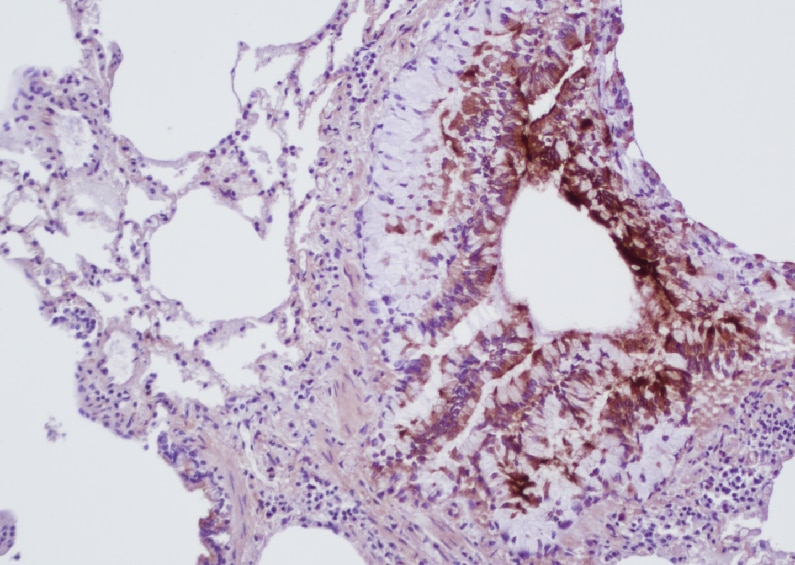
 fibrosis, B: Non-fibrotic CV, C: Perisinusodial fibrosis, D: Non-fibrotic area, E: Protat tract fibrosis, F: Septal fibrosis (arrow). Antigen retrieval: not required. Primary antibody: Anti-collagen type I at 1:1250 for 4C for 24hr. Secondary antibody: Peroxidase biotin-streptavidin rabbit secondary antibody at 1:10,000 for 45 min at RT. Localization: Anti-collagen type I is intra and extracellular. Staining: 3.3'-diaminobenzidine tetrahydrochloride was used as the chromogen. Nuclei were counterstained purple with hematoxylin.")


 with hematoxylin purple nuclear counterstain. With corresponding negative conrol (B).")

, licensed under a CC-BY license.")


 fibrosis, B: Non-fibrotic CV, C: Perisinusodial fibrosis, D: Non-fibrotic area, E: Protat tract fibrosis, F: Septal fibrosis (arrow). Fixation: formalin fixed paraffin embedded. Antigen retrieval: not required. Primary antibody: Anti-collagen type I at 1:1250 for 4C for 24hr. Secondary antibody: Peroxidase biotin-streptavidin rabbit secondary antibody at 1:10,000 for 45 min at RT. Localization: Anti-collagen type I is intra and extracellular. Staining: 3.3'-diaminobenzidine tetrahydrochloride was used as the chromogen. Nuclei were counterstained purple with hematoxylin.")
![Immunohistochemistry-Paraffin: Rabbit Polyclonal Collagen I Antibody [NB600-408] - Collagen I was stained in human FFPE tissue. Primary antibody dilution: 1/1000 in blocking buffer. HIER antigen retrieval at pH 9 for 20min. AF750 conjugated version of the antibody was used (Catalog # NB600-408AF750). Image from a verified customer review.](http://images.novusbio.com/fullsize/antibody/nb600-408_rabbit-polyclonal-collagen-i-antibody-immunohistochemistry-paraffin-1122023154839..jpg "Immunohistochemistry-Paraffin: Rabbit Polyclonal Collagen I Antibody [NB600-408] - Collagen I was stained in human FFPE tissue. Primary antibody dilution: 1/1000 in blocking buffer. HIER antigen retrieval at pH 9 for 20min. AF750 conjugated version of the antibody was used (Catalog # NB600-408AF750). Image from a verified customer review.")
 Pancreatic tumor tissues from vehicle, gemcitabine, or combination of gemcitabine plus fasudil treated KPC mice were harvested and stained for various stromal markers. Representative images are shown of H&E staining, alpha -SMA, Desmin, CD31, Collagen I, and Movat's pentachrome staining.")
 and collagen 2 (D and E) among the anterior horn, the body and the posterior horn: (A and D) young model; (B and E) adult model. Comparison of collagen 1 (C) and collagen 2 (F) content between the young and the adult specimens. Values with different superscripts differ for P < 0.01 (A,B). (G) Representative Western blot image for collagen 1, collagen 2 and GAPDH. N/group = 8.")
![EPS15 is targeted via a SPOP/SPOPL binding consensus motif.(A) Cartoon of human EPS15 domain-organization and the amino-acid sequence. Indicated by color code are the SPOP/SPOPL binding site (red) and the lysine residue (yellow), which is ubiquitinated in a CRL3SPOPL–dependent manner in vivo. In addition, the amino-terminal Ca2+-binding EF-hand motifs (EH), the coiled-coil domain involved in dimerization and the two carboxy-terminal ubiquitin-interacting motifs (UIMs) involved in ubiquitin-binding are indicated. (B) EPS15 ubiquitin-profiling. Peptides containing EPS15 modification sites were quantified with LC-MS/MS after enrichment of the K-epsilon -GG motif from whole cell digests of HeLa cells treated with siSPOPL or siControl. Normalized precursor mass intensity profiles for EPS15 sites corresponding to K793, K801 and K693 are shown (raw data in Figure 4—figure supplement 1B). Quantification of the beta -Actin K113 and the polyubiquitin K11 linkage peptide control for comparable enrichment. Data are mean ± SD, N = 3. **p≤0.01. (C) Purified SPOPL was incubated as indicated with GST-tagged wild-type EPS15 or GST-EPS15 mutants, where the predicted SPOPL binding motifs have been mutated individually (GST-EPS15S605-S607A and EPS15S744-S746A, respectively), pulled down with glutathione sepharose (IP [GST]) and bound proteins were analyzed by Coomassie blue staining (upper panel) and immunoblotting (lower panels). Note that SPOPL readily binds to GST-EPS15 and GST-EPS15S605-S607A, but this interaction is strongly reduced with the GST-EPS15S744-S746A mutant. (D) HeLa cells stably expressing GFP-tagged wild-type EPS15, the EPS15S744-S746A or the EPS15K793R mutants from a doxycycline-inducible promoter were transfected as indicated (+) with control siRNA or siRNA depleting SPOPL. The levels of EPS15-GFP, EGFR and for control tubulin (TUB) were analyzed by immunoblotting with specific antibodies. Experiments were quantified in Fiji and the EPS15 levels plotted as fold-increase compared to controls. Data are mean ± SEM, N = 4. *p≤0.05. Note that SPOPL depletion does not further increase the levels of both EPS15 mutants. (E) Total cell extracts were prepared from HeLa cells expressing either GFP-tagged wild-type, the EPS15S744-S746A mutant or the EPS15K793R mutant in the presence (+) or absence (-) of HA-tagged SPOPL overexpression. The levels of EPS15-GFP, SPOPL-HA and control GAPDH were analyzed by immunoblotting. Note that overexpression of SPOPL-HA is able to induce degradation of wild-type but not the EPS15S744-S746A-GFP or the EPS15K793R-GFP mutant.DOI://dx.doi.org/10.7554/eLife.13841.009EPS15 is targeted via a SPOP/SPOPL binding consensus motif.(A) Alignments of the carboxy-terminal domains of EPS15 proteins from various species. Conserved SPOPL-binding motifs and putative ubiquitination sites are highlighted by yellow boxes. (B) Peptides containing EPS15 modification sites were quantified with LC-MS/MS after enrichment of the K-epsilon -GG motif from whole cell HeLa digests treated with siSPOPL and siControl. Raw intensities for each of the triplicate LC-MS/MS runs are shown with each of the siControl conditions scaled to 100% intensity. Normalized precursor mass intensity profiles for EPS15 sites corresponding to K793, K801 and K693 are shown, with only K793 showing significant downregulation in the depletion condition. Quantification of a peptide corresponding to beta -Actin K113 and the poly-ubiquitin K11 linkage peptide is also shown to demonstrate that enrichment variations did not influence the quantification of the EPS15 sites. Additionally, the total ion chromatographic intensities for each run are plotted to provide insight into the consistency of each of the separate experiments performed on different days. Data are mean ± SD, N = 3. (C) HeLa cell lines stably expressing wild-type EPS15-GFP, the EPS15S744-746A-GFP mutant or the EPS15K793R-GFP mutant from the inducible doxycycline-promoter were treated with doxycycline for 3 days, and analyzed by live cell imaging. Displayed are maximal projections of Z-stack acquisitions, fully covering cell height. Scale bar = 10 μm.DOI://dx.doi.org/10.7554/eLife.13841.010 Image collected and cropped by CiteAb from the following open publication (//elifesciences.org/articles/13841), licensed under a CC-BY license. Not internally tested by Novus Biologicals.](http://images.novusbio.com/fullsize/nb600-408_rabbit-collagen-i-pab-23220248194310.jpg "EPS15 is targeted via a SPOP/SPOPL binding consensus motif.(A) Cartoon of human EPS15 domain-organization and the amino-acid sequence. Indicated by color code are the SPOP/SPOPL binding site (red) and the lysine residue (yellow), which is ubiquitinated in a CRL3SPOPL–dependent manner in vivo. In addition, the amino-terminal Ca2+-binding EF-hand motifs (EH), the coiled-coil domain involved in dimerization and the two carboxy-terminal ubiquitin-interacting motifs (UIMs) involved in ubiquitin-binding are indicated. (B) EPS15 ubiquitin-profiling. Peptides containing EPS15 modification sites were quantified with LC-MS/MS after enrichment of the K-epsilon -GG motif from whole cell digests of HeLa cells treated with siSPOPL or siControl. Normalized precursor mass intensity profiles for EPS15 sites corresponding to K793, K801 and K693 are shown (raw data in Figure 4—figure supplement 1B). Quantification of the beta -Actin K113 and the polyubiquitin K11 linkage peptide control for comparable enrichment. Data are mean ± SD, N = 3. **p≤0.01. (C) Purified SPOPL was incubated as indicated with GST-tagged wild-type EPS15 or GST-EPS15 mutants, where the predicted SPOPL binding motifs have been mutated individually (GST-EPS15S605-S607A and EPS15S744-S746A, respectively), pulled down with glutathione sepharose (IP [GST]) and bound proteins were analyzed by Coomassie blue staining (upper panel) and immunoblotting (lower panels). Note that SPOPL readily binds to GST-EPS15 and GST-EPS15S605-S607A, but this interaction is strongly reduced with the GST-EPS15S744-S746A mutant. (D) HeLa cells stably expressing GFP-tagged wild-type EPS15, the EPS15S744-S746A or the EPS15K793R mutants from a doxycycline-inducible promoter were transfected as indicated (+) with control siRNA or siRNA depleting SPOPL. The levels of EPS15-GFP, EGFR and for control tubulin (TUB) were analyzed by immunoblotting with specific antibodies. Experiments were quantified in Fiji and the EPS15 levels plotted as fold-increase compared to controls. Data are mean ± SEM, N = 4. *p≤0.05. Note that SPOPL depletion does not further increase the levels of both EPS15 mutants. (E) Total cell extracts were prepared from HeLa cells expressing either GFP-tagged wild-type, the EPS15S744-S746A mutant or the EPS15K793R mutant in the presence (+) or absence (-) of HA-tagged SPOPL overexpression. The levels of EPS15-GFP, SPOPL-HA and control GAPDH were analyzed by immunoblotting. Note that overexpression of SPOPL-HA is able to induce degradation of wild-type but not the EPS15S744-S746A-GFP or the EPS15K793R-GFP mutant.DOI://dx.doi.org/10.7554/eLife.13841.009EPS15 is targeted via a SPOP/SPOPL binding consensus motif.(A) Alignments of the carboxy-terminal domains of EPS15 proteins from various species. Conserved SPOPL-binding motifs and putative ubiquitination sites are highlighted by yellow boxes. (B) Peptides containing EPS15 modification sites were quantified with LC-MS/MS after enrichment of the K-epsilon -GG motif from whole cell HeLa digests treated with siSPOPL and siControl. Raw intensities for each of the triplicate LC-MS/MS runs are shown with each of the siControl conditions scaled to 100% intensity. Normalized precursor mass intensity profiles for EPS15 sites corresponding to K793, K801 and K693 are shown, with only K793 showing significant downregulation in the depletion condition. Quantification of a peptide corresponding to beta -Actin K113 and the poly-ubiquitin K11 linkage peptide is also shown to demonstrate that enrichment variations did not influence the quantification of the EPS15 sites. Additionally, the total ion chromatographic intensities for each run are plotted to provide insight into the consistency of each of the separate experiments performed on different days. Data are mean ± SD, N = 3. (C) HeLa cell lines stably expressing wild-type EPS15-GFP, the EPS15S744-746A-GFP mutant or the EPS15K793R-GFP mutant from the inducible doxycycline-promoter were treated with doxycycline for 3 days, and analyzed by live cell imaging. Displayed are maximal projections of Z-stack acquisitions, fully covering cell height. Scale bar = 10 μm.DOI://dx.doi.org/10.7554/eLife.13841.010 Image collected and cropped by CiteAb from the following open publication (//elifesciences.org/articles/13841), licensed under a CC-BY license. Not internally tested by Novus Biologicals.")


![N/A PSG1 [HRP]](https://images.novusbio.com/images2/DATA_PSG1_DPSG10_ELISA_797.jpg)
![N/A PSG1 [HRP]](https://images.novusbio.com/images2/PSG-1_DPSG10_ELISA_164.jpg)
![COCO/DAND5 [Unconjugated]](/sites/all/modules/enterprise-tech/et_datasheets/images/novus_guarantee.png "COCO/DAND5 [Unconjugated]")
![Immunocytochemistry RUNX2/CBFA1 Antibody (232902) [Unconjugated]](https://images.novusbio.com/images2/RUNX2_MAB2006_Immunocytochemistry_13309.jpg)


![N/A IL-6 [HRP]](https://images.novusbio.com/images2/DATA_IL6_M6000_ELISA_936.jpg)
![N/A IL-6 [HRP]](https://images.novusbio.com/images2/IL-6_M6000_ELISA_415.jpg)
![N/A IL-6 [HRP]](https://images.novusbio.com/images/m6000b_mouse-il-6-quantikine-elisa-kit-44202415433118.jpg)
![SDS-PAGE PDGF-BB [Unconjugated]](https://images.novusbio.com/images2/PDGF-BB_220-BB_290.jpg)
![Bioactivity PDGF-BB [Unconjugated]](https://images.novusbio.com/images2/PDGF-BB_220-BB_291.jpg)
![Immunohistochemistry CTGF/CCN2 Antibody [Unconjugated] - C-terminus](https://images.novusbio.com/images2/CTGF_AF660_Immunohistochemistry_9968.jpg)
![Immunohistochemistry CTGF/CCN2 Antibody [Unconjugated] - C-terminus](https://images.novusbio.com/images2/CTGF_AF660_Immunohistochemistry_9969.jpg)
![Western Blot CTGF/CCN2 Antibody [Unconjugated] - C-terminus](https://images.novusbio.com/images/af660_human-ctgf-ccn2-c-terminus-affinity-purified-polyclonal-ab-822024951141.jpg)
![Bioactivity TGF-beta 1 [Unconjugated]](https://images.novusbio.com/images/protein/7754-bhcf_recombinant-human-tgf-beta-1-human-cell-expressed-cf-bioactivity-1811202013946.jpg)
![Bioactivity BMP-2 [Unconjugated]](https://images.novusbio.com/images/protein/355-bm_recombinant-human-mouse-rat-bmp-2-protein-bioactivity-181120209742.jpg)
![SDS-PAGE IGF-I/IGF-1 [Unconjugated]](https://images.novusbio.com/images2/IGF-I_291-G1_40.jpg)
![Bioactivity IGF-I/IGF-1 [Unconjugated]](https://images.novusbio.com/images2/IGF-I_291-G1_41.jpg)
![Mass Spectrometry IGF-I/IGF-1 [Unconjugated]](https://images.novusbio.com/images2/IGF-I_291-G1_42.jpg)
 using a 1:1000 dilution of HRP-conjugated Anti-Rabbit IgG Secondary Antibody (Catalog # HAF008). This experiment was conducted under reducing conditions and using Immunoblot Buffer Group 1.")
![Western Blot: Goat anti-Rabbit IgG (H+L) Secondary Antibody [HRP] [NB7160] - Western blot showing vemurafenib treatment in BRAFV600E CRC cells inhibits fission mediator DRP1 with no significant effect on fusion proteins (Mfn1 & 2) using MFN-1 antibody (NBP1-51841) and corresponding secondary antibody, goat anti-rabbit IgG-HRP (NB7160). Image collected and cropped by CiteAb from the following publication (https://pubmed.ncbi.nlm.nih.gov/33738242).](https://images.novusbio.com/images/Goat-anti-Rabbit-IgG-H+L-Secondary-Antibody-HRP-Western-Blot-NB7160-img0001.jpg "Western Blot: Goat anti-Rabbit IgG (H+L) Secondary Antibody [HRP] [NB7160] - Western blot showing vemurafenib treatment in BRAFV600E CRC cells inhibits fission mediator DRP1 with no significant effect on fusion proteins (Mfn1 & 2) using MFN-1 antibody (NBP1-51841) and corresponding secondary antibody, goat anti-rabbit IgG-HRP (NB7160). Image collected and cropped by CiteAb from the following publication (https://pubmed.ncbi.nlm.nih.gov/33738242).")
![Flow Cytometry: Rabbit IgG Isotype Control [NBP2-24891] - Intracellular FACS analysis of mouse TLR6 polyclonal antibody (red), rabbit isotype control (green), RAW cells alone (shaded). Two micrograms of antibodies were used. Goat anti-rabbit FITC (Novus, 20302) was used as secondary.](https://images.novusbio.com/images/Rabbit--Mouse-IgG-Isotype-Control-Flow-Cytometry-NBP2-24891-img0001.jpg "Flow Cytometry: Rabbit IgG Isotype Control [NBP2-24891] - Intracellular FACS analysis of mouse TLR6 polyclonal antibody (red), rabbit isotype control (green), RAW cells alone (shaded). Two micrograms of antibodies were used. Goat anti-rabbit FITC (Novus, 20302) was used as secondary.")